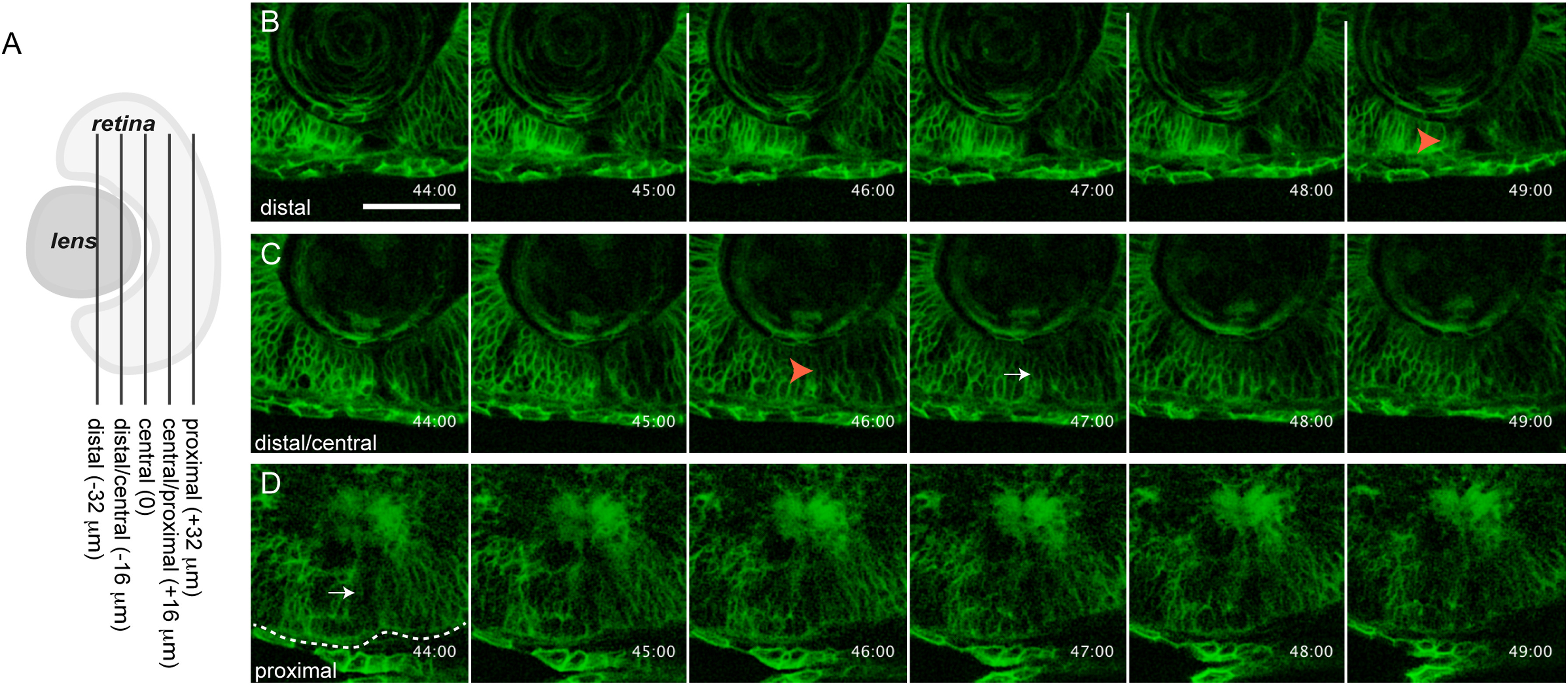
Fig. 2
in vivo imaging of choroid fissure closure in zebrafish. (A) Schematic depicting the approximate level of sections in B-D along the proximal-distal axis of the CF. The vitreous cavity was defined as central, and optical sections were taken at 16 Ám intervals proximally and distally from this point. (B-D) membrane-GFP injected embryos were imaged throughout the CF. Single micron optical slices are shown from 44 to 49 hpf and at three distinct proximal-distal regions of the CF. (B) Distally, the CF remains open until at least 49 hpf. (C) Distal/centrally, the CF appears to close between 46?47 hpf. (D) Proximally, the CF already appears to be closed at 44 hpf. Orange arrowheads in B,C mark open CF. White arrow in C marks what appears to be a closed CF. Dashed line outlines the RPE. Scale bar=50 Ám.
Reprinted from Developmental Biology, 419(2), James, A., Lee, C., Williams, A.M., Angileri, K., Lathrop, K.L., Gross, J.M., The Hyaloid Vasculature Facilitates Basement Membrane Breakdown During Choroid Fissure Closure in the Zebrafish Eye, 262-272, Copyright (2016) with permission from Elsevier. Full text @ Dev. Biol.

