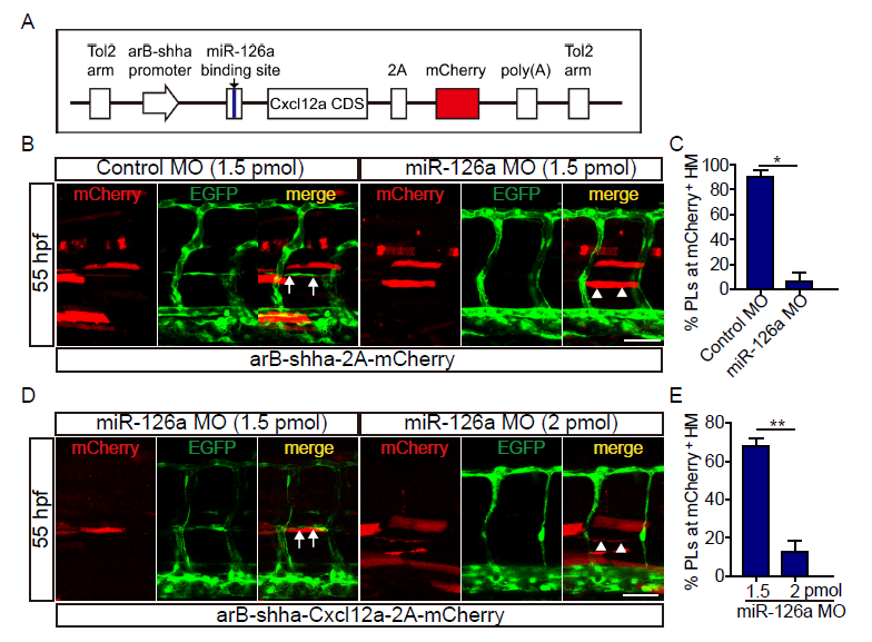
Fig. S9
Cxcl12a re-expression could not rebuild the trunk lymphatic vessels under miR-126a?s dramatic knockdown. (A) Diagram of Cxcl12a rescue constructs for spatial overexpression of Cxcl12a accompanied with mCherry. (B) Confocal images of partial trunk vessels at 55 hpf embryos. White arrows indicate normally formed PL colocalization with Cxcl12a and white arrowheads denote absence of PL. Scale bar, 50 ?m. (C) Quantification of the percentage of PL formed at mCherry positive HM at 55 hpf. Compared with 1.5 pmol control MO group, the PL in 1.5 pmol miR-126a MO group was severely absent upon mCherry expression. *p<0.05. (D) Confocal images of partial trunk vessels at 55 hpf embryos. White arrows indicate reformed PL colocalization with Cxcl12a and white arrowheads denote absence of PL. Scale bar, 50 ?m. (E) Quantification of the percentage of PL formed at mCherry positive HM at 55 hpf. Compared with 1.5 pmol MO knockdown group, the PL in 2 pmol MO group was more severely absent upon Cxcl12a re-expression. **p<0.01.

