
|
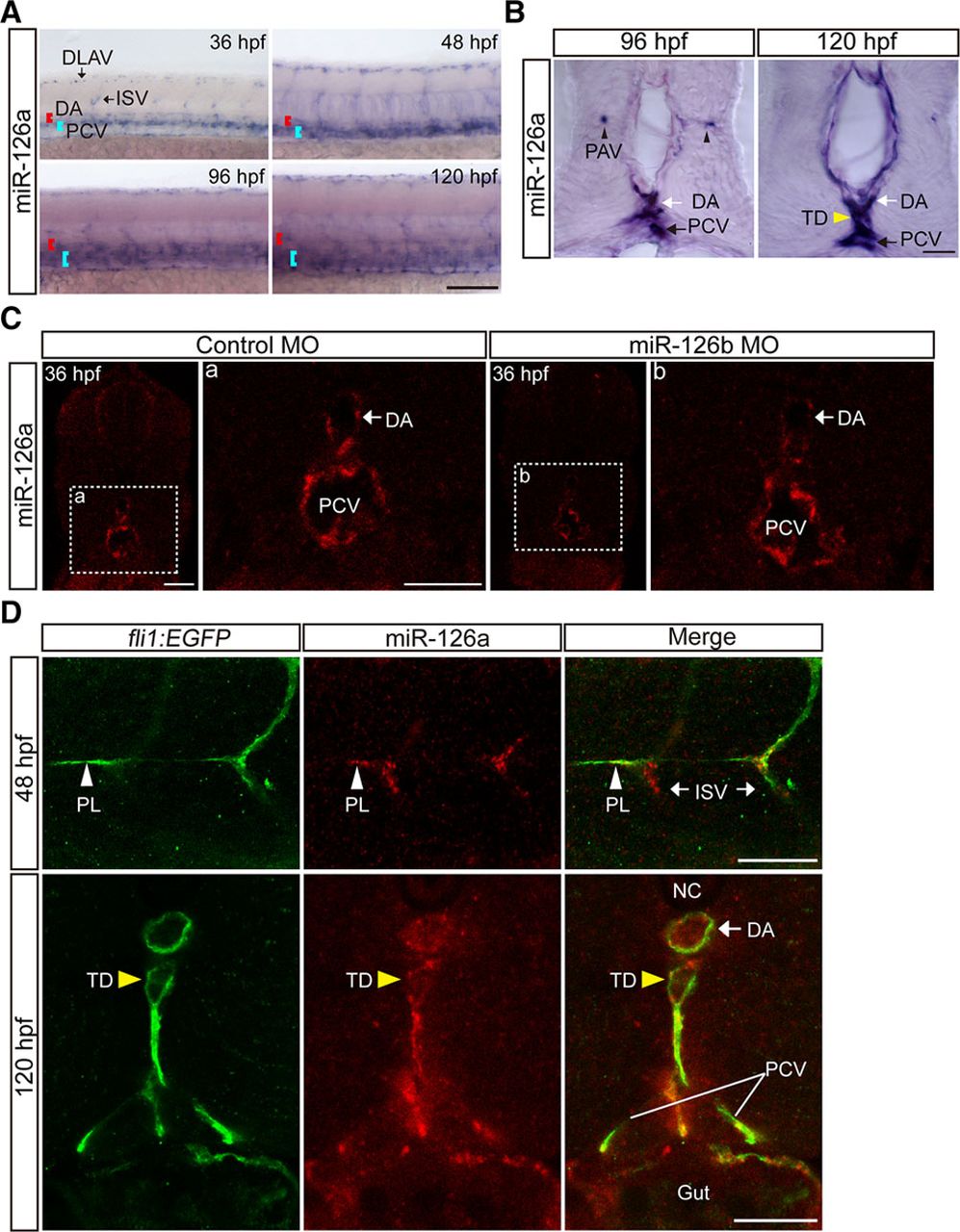
Fig. 2
MiR-126a is expressed in lymphatic vessels. A, Whole-mount in situ hybridization (WISH) for miR-126a at different stage embryos. The head of embryos toward the left and the dorsal sides of the embryo are at the top of the figures. Scale bar, 200 ?m. B, Cross-sectional images of miR-126a stained by WISH. Scale bars, 10 ?m. C, Cross-sectional images of miR-126a stained by fluorescent in situ hybridization (FISH). Scale bars, 25 ?m. D, Top, Trunk of miR-126bKD embryo at 48 hours post fertilization (hpf), stained by immunofluorescence (IF) for enhanced green fluorescent protein (EGFP) and by FISH for miR-126a. Bottom, Transverse sections through the trunk of a 120 hpf miR-126bKD embryo, stained by the same staining procedure as (Top). Scale bars, 25 ?m. DA indicates dorsal aorta; DLAV, dorsal longitudinal anatomic vessel; ISV, intersegmental vessel; MO, morpholino; NC, Notochord; PAV, parachordal vessel; PCV, posterior cardinal vein; PL, parachordal lymphangioblast; and TD, thoracic duct.