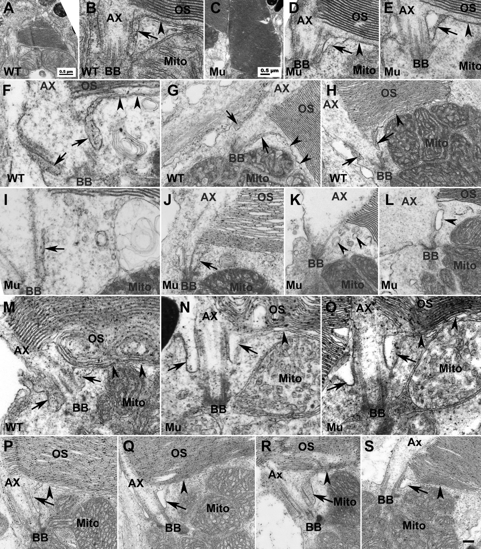
Fig. 3
EYS deficiency caused disruption of the ciliary pocket in cones. To determine the impact of EYS deficiency on structures near CC/TZ, we performed EM analysis on EYS-deficient zebrafish retina at 7, 40, 50, 80?dpf and 8 and 14?mpf. (A,B) Low and high magnification of same wild-type cone photoreceptor at 7?dpf. (C,D) Low and high magnification of the same EYS-deficient cone photoreceptor at 7?dpf. (E) Another example of EYS-deficient cone photoreceptor at 7?dpf. (F-H) Wild-type cone photoreceptors at 40?dpf. (I-L) EYS-deficient zebrafish cone photoreceptors at 40?dpf. (M) Wild-type rod photoreceptor at 40?dpf. (N,O) EYS-deficient rod photoreceptors at 40?dpf. (P) Wild-type rod photoreceptor at 8?mpf. (Q) EYS-deficient rod photoreceptor at 8?mpf. (R) Wild-type rod photoreceptor at 14?mpf. (S) EYS-deficient rod photoreceptor at 14?mpf. In wild type at 7 and 40?dpf cones, the ciliary pocket with electron dense lumen was clearly observed (B,F-H, arrows). The plasma membrane separating the outer and inner segments was in continuity with the membrane of the ciliary pocket (arrowheads in B,F-H). In EYS-deficient animals at 7?dpf, the cone ciliary pocket was normal except that the lumen of the mutant ciliary pocket was not as electron dense as the wild type (D,E, arrows). At 40?dpf, in EYS-deficient animals, the ciliary pocket in cone photoreceptors was collapsed (I and J, arrows) or was replaced with multiple membrane vesicles of electron density (K,L, arrowheads). The rod ciliary pocket in EYS-deficient zebrafish was largely maintained (M-O,Q,S, arrows). Scale bar in S: 100?nm for B,D-F,I,M-O; 200?nm for G,H,J,L,P-S. Ax, axoneme; BB, basal body; Mito, mitochondria; OS, outer segment.

