
Fig. 8
|
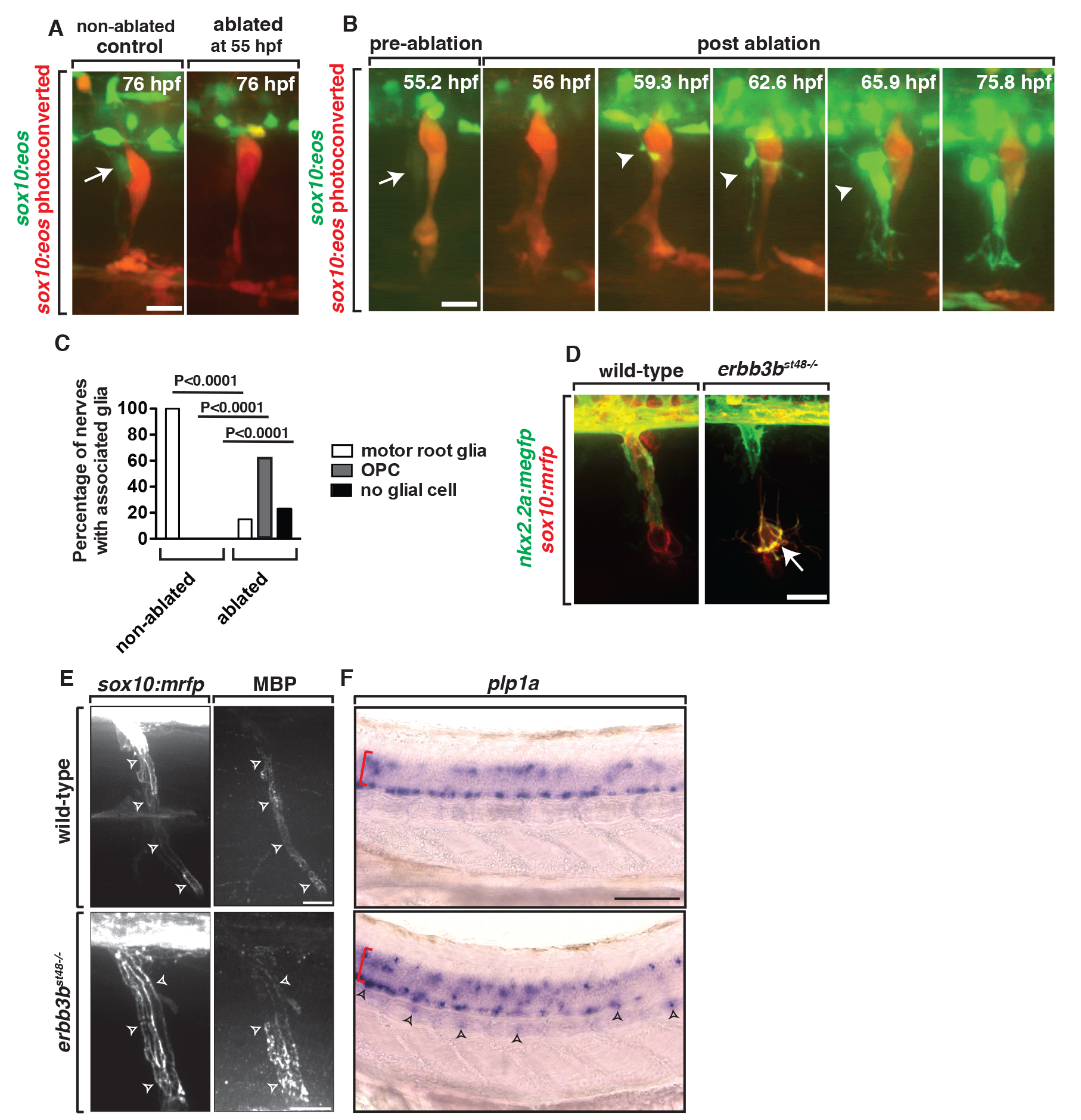
Fig. 8
Ablation of MEP glia disrupts the MEP TZ.
(A) In a control Tg(sox10:eos) embryo that was exposed to UV light at 48 hpf, motor (green) and sensory root (red) glial cells ensheath axons. When the CNS-derived MEP glial cell progenitor was ablated at 55 hpf and then imaged at 76 hpf, all sox10+ motor root glial cells were absent. (B) Frames captured from a 16-h time-lapse video beginning at 56 hpf in a Tg(sox10:eos) embryo exposed to UV light at 48 hpf. Numbers in upper right corners denote stage of embryos. When the MEP glial cell progenitor (arrow) was ablated at 55 hpf, OPC processes (arrowhead) extended out of the spinal cord before OPC cell bodies exited. All images are lateral views of the motor and sensory root with dorsal to the top and anterior to the left. (C) Quantification of the data in panel B (n?=?46 nerves). (D) In Tg(nkx2.2a:mgfp);Tg(sox10:mrfp);erbb3b?/? embryos at 55 hpf, motor root glial cells are absent and OPCs (arrows) are in the PNS. (E) In Tg(sox10:mrfp) larva at 8 dpf stained with MBP, MBP+ cells were ensheathed around the root in wild-type and erbb3 animals. (F) In situ hybridization of plp1a in wild-type and erbb3b mutant showed OPCs in the PNS in erbb3b mutants myelinate with CNS myelin. Scale bars, 25 �m.