Fig. 6
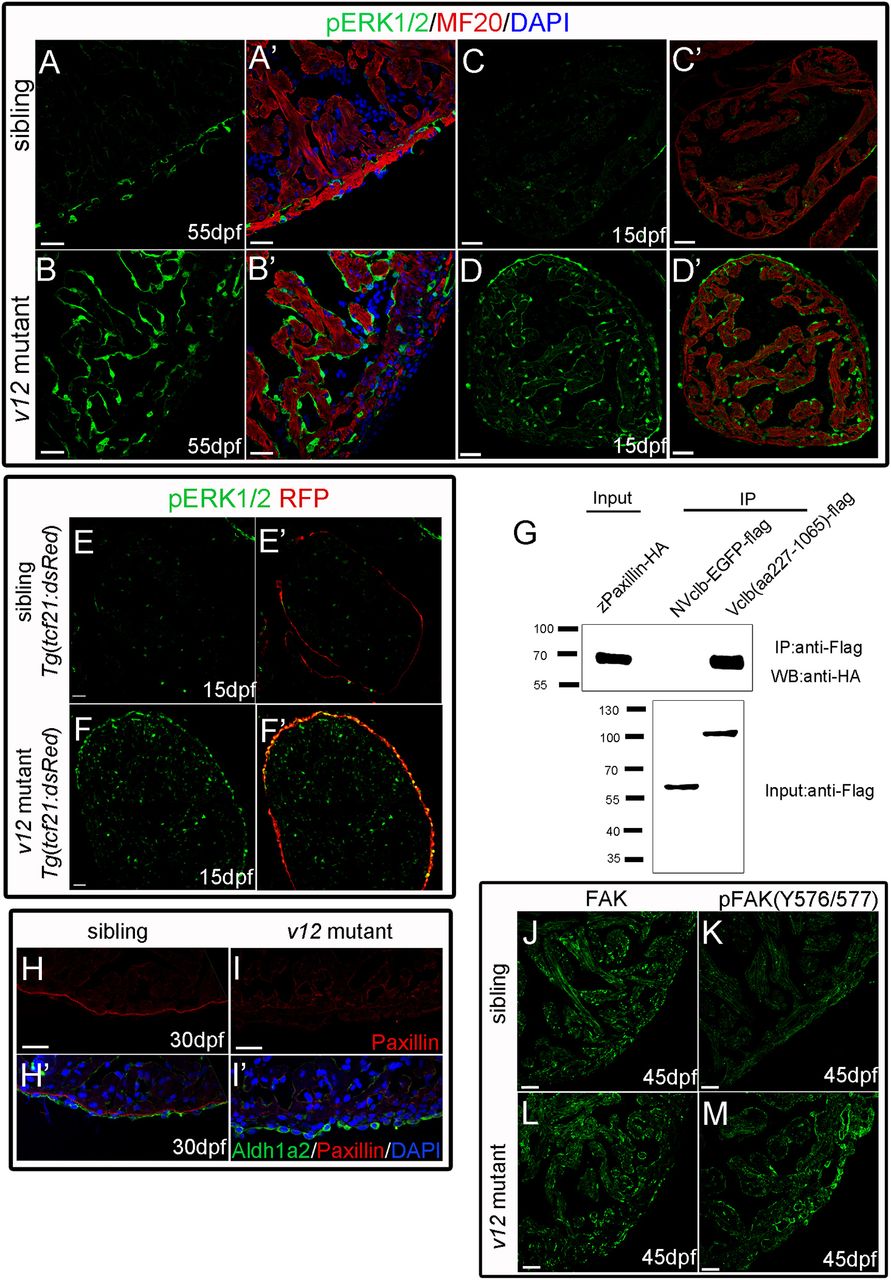
pERK and pFAK are upregulated in the epicardium and endocardium of v12 mutant ventricles. (A-B') pErk1/2 expression (green) is increased in the endocardium of v12 mutants at 55?dpf compared with wild-type sibling. (C-D') pErk1/2 expression is increased in the epicardium and endocardium of v12 mutant at 15?dpf. MF20 staining (red) indicates cardiac muscles. (E-F') Tg(tcf21:dsRed) transgenic reporter indicates epicardium. Upregulated pErk1/2 colocalizes with RFP signal. (G) Co-immunoprecipitation shows that NVclb-EGFP is not associated with paxillin, which normally interacts with wild-type Vclb, when ectopically expressed in HEK293 cells. IP, immunoprecipitation; WB, western blot. Size markers (kDa) are shown to the left. (H-I') The localization of paxillin at the boundary between epicardium and myocardium is partially lost in the v12 mutant (L,L') compared with wild-type siblings (H,H'). (J-M) pFAK(Y576/577) expression is upregulated whereas total FAK expression remains unchanged in the epicardium and endocardium of the v12 mutant at 45?dpf (L,M) compared with wild-type sibling (J,K). Scale bars: 20??m.

