Fig. 3
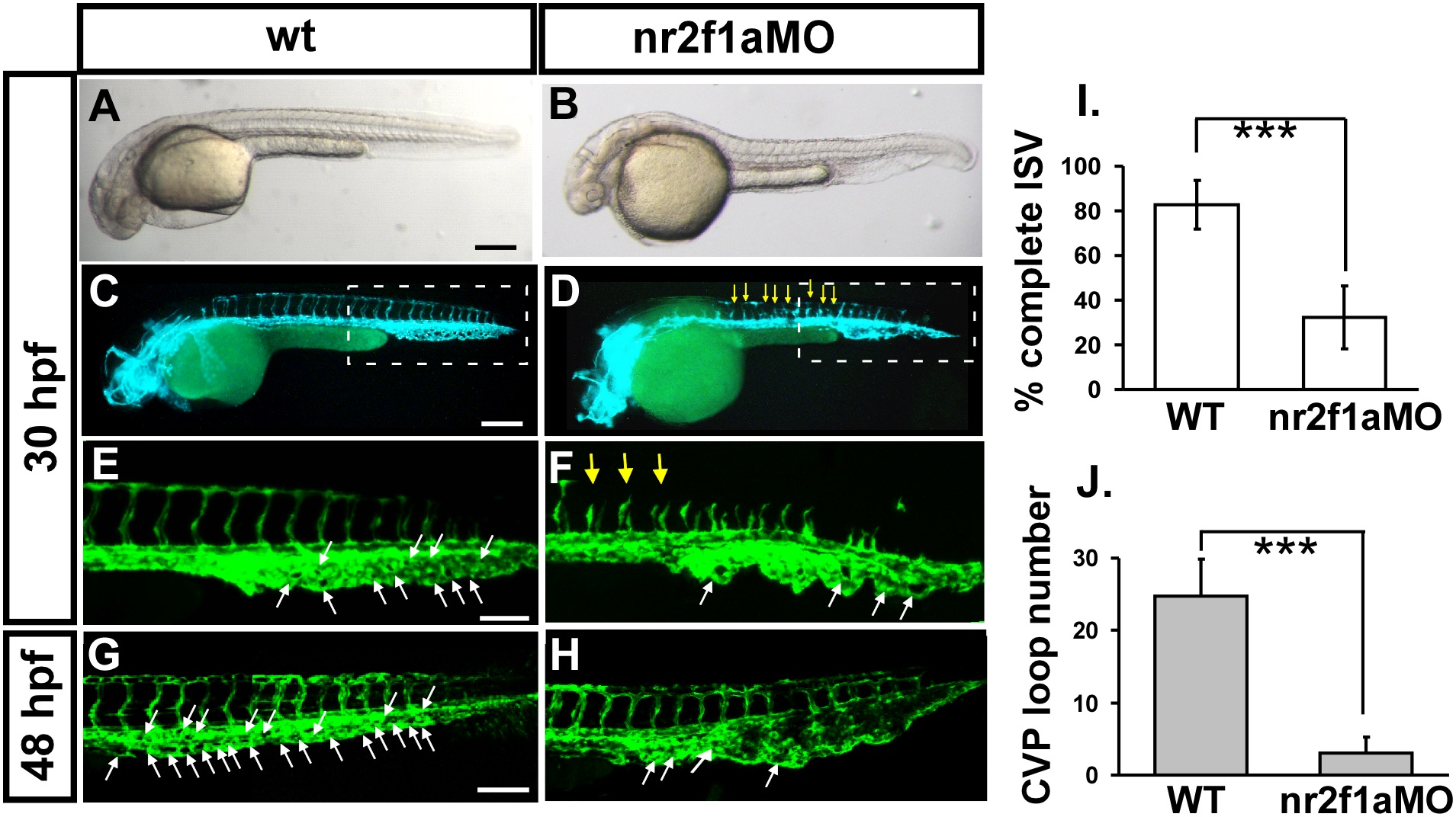
Knockdown of nr2f1a causes defects in zebrafish vascular development.
(A-H) Loss of nr2f1a showing ISV growth defect (yellow arrows in F) and mis-pattern plexus at the CVP (caudal vein plexus) (white arrows) compared to wild-type control (E) at 30 hpf. E and F is enlargement of C and D, respectively. At 48 hpf, the swallowed plexus becomes obvious (H). At 30 hpf, in uninjected control embryos, intersegmental vessels (isv) have reached the DLAV at the dorsal aspect of the embryo (C, E) and the caudal vein plexus (cvp) formed honeycomb-like structures at the tail (E, white arrows). At the same stage ISVs are stalled at mid-somite in nr2f1ae2i2 morphants (D, F). (I) Quantification of percentage of completed ISV shows a ~50% increase compared to nr2f1a morphants (n = 26 in wt and n = 34 in nr2f1aMO) at 30 hpf. (J) Quantification of loop formation at CVP shows a 4-fold decreased in nr2f1a morphants (n = 10 in wt and in nr2f1aMO) at 48 hpf. (*** refers to p<0.0001 by an unpaired student′s t-test. Scale bars are 200 µm for A-D, and 100 µm for E-H.

