Fig. 1
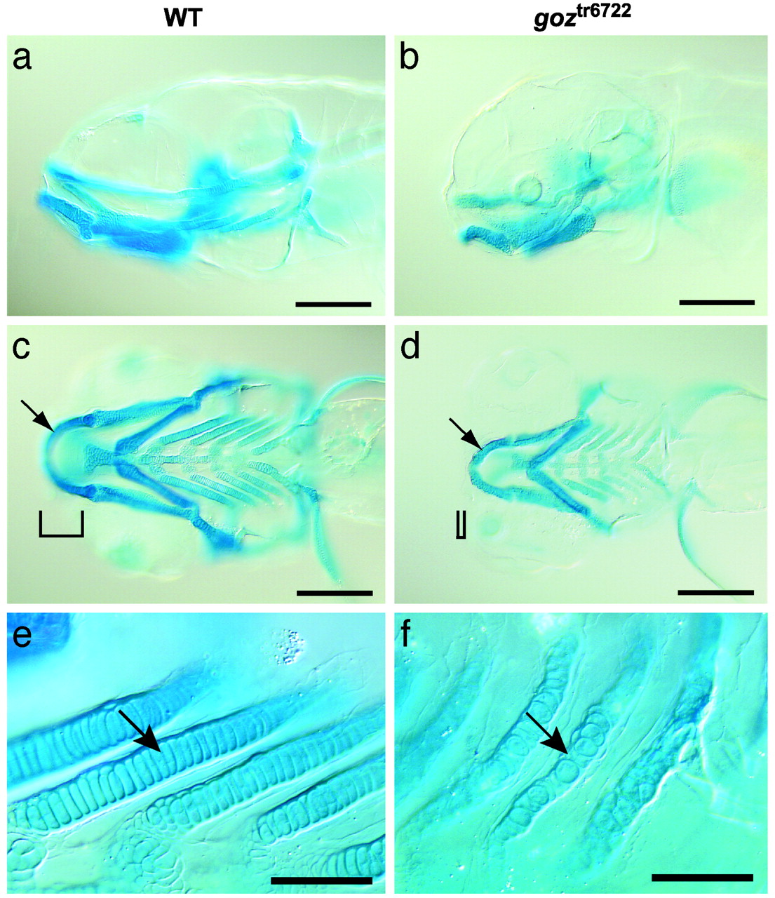
Cartilage defects in goz. Alcian blue staining of sibling larvae (a, c, and e) and goz larvae (b, d, and f) at 5 dpf is shown. (a and b) Lateral view. (c and d) Ventral view of the head. (e and f) Enlargement of the branchial arches. Cartilage matrix staining is slightly reduced in goz mutants. All cartilage elements, including Meckel′s cartilage (arrows in c and d), are smaller in the mutant. Tissue anterior to the eyes is missing (bars in c and d). The columnar arrangement of chondrocytes in the branchial arches is disrupted in goz mutants (arrows in e and f). (Scale bars are 200 Ámin a-d and 50 Ámin e and f.)

