
Image
|
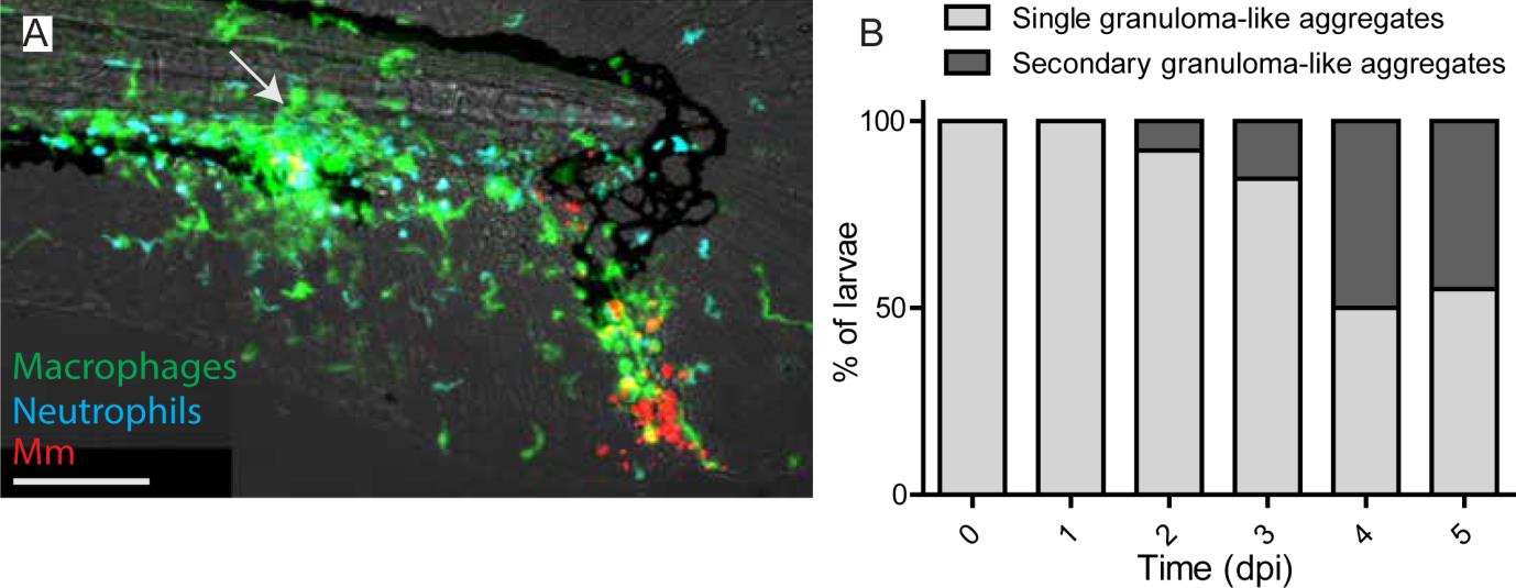
Figure Caption
Fig. S3
Secondary granulomas at late stages of infection. A) Representative image of an infected tail fin showing a secondary granuloma like aggregate (arrow) near the caudal vein. B) Percentage of larvae showing formation of a secondary granuloma like aggregate at different stages of infection. Scale bar: 100 �m.
Acknowledgments
This image is the copyrighted work of the attributed author or publisher, and
ZFIN has permission only to display this image to its users.
Additional permissions should be obtained from the applicable author or publisher of the image.
Full text @ J. Cell Sci.