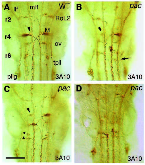
Fig. 8
Variable disorganization of hindbrain reticulospinal interneurons in pactj250a at 36 hpf shown by 3A10 mAb staining; dorsal views with rostral to the top. In wild type (A), the most prominently labeled Mauthner cells are located in r4, which project axons contralaterally to the mlf. RoL2 exists in r2, which projects axons contralaterally to the mlf, then to the llf. In A, B and C, MiM1(or MiV1) (arrowheads; Mendelson, 1986a, b) is indicated. According to their position and axonal projection, the two neurons in r6 could be MiD3i (asterisk), which has an ipsilateral axon and MiD3c (triangle), which has a contralateral axon, as shown in C. B, C and D are representatively labeled embryos. In most of the cases, as shown in B, one of the Mauthner cells in rhombomere 4 is displaced to a more posterior rhombomere (r5, r6 or in between) and locates in the same side of the normal one (n=5/10). In B, a presumptively displaced MiD3c (arrow) is indicated. In a few cases, as shown in C, the displaced Mauthner cell locates in the opposite side (n=1/10). Sometimes, as shown in D, both of the Mauthner cells are displaced to a more posterior rhombomere, and their axons project to the ipsilateral instead of the contralateral side (n=3/10). llf, lateral longitudinal fascicle; M, Mauthner cell; mlf, medial longitudinal fascicle; ov, otic vesicle; pllg, posterior lateral line ganglion; RoL2, RoL2 neurons in r2; tpll, tract of posterior lateral ganglion. Bar, 125 Ám.

