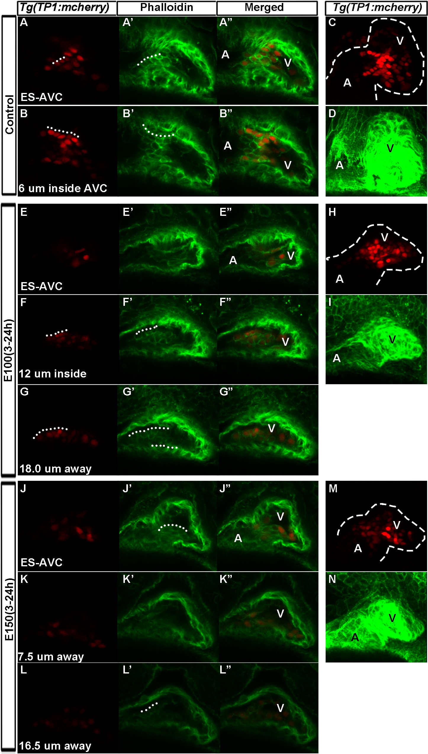
Fig. 8
High Notch-active cells present in the ventricular endocardium in ethanol exposed embryos showed F-actin expression.
(A-B′′) Texas-red phalloidin stained 52 hpf Tg(TP1:mCherry) embryos showed strong mCherry labelled endocardial cells confined at the AVC in control embryo (A-A′′: at the surface of AVC; (B-B′′): cells inside AVC); Strong Notch positive cells along with a few neighboring cells expressed high level of F-actin and were cuboidal in shape (White dotted lines). (C) 3D rendering of optical Z-sections through heart showing distribution of strong and weak Notch active cells in the heart of control embryo. (D) 3D rendering of phalloidin stained embryos showing shape of the control embryo heart. (E-G′′, J-L′′) Ethanol treated and Texas-red phalloidin stained Tg(TP1:mCherry) embryos showed strong mCherry labelled endocardial cells at a lesser extent at the AVC endocardium (E-E′′, J-J′′), but more inside the ventricle (F-G′′, K-L′′). Notch active cells in the chamber endocardium showed ectopic F-actin labeling (F′-G′′, K′-L′′). White dotted lines show cuboidal cells. (H, M) 3D rendering of optical Z-sections through heart showing distribution of strong and weak Notch active cells in the heart of ethanol treated embryos. (I, N) 3D rendering phalloidin stained embryos showing the shape of the heart of ethanol treated embryos compared to control (D). ES-AVC: Endocardial cells at the surface of AVC, A: Atrium, V: Ventricle.

