
|
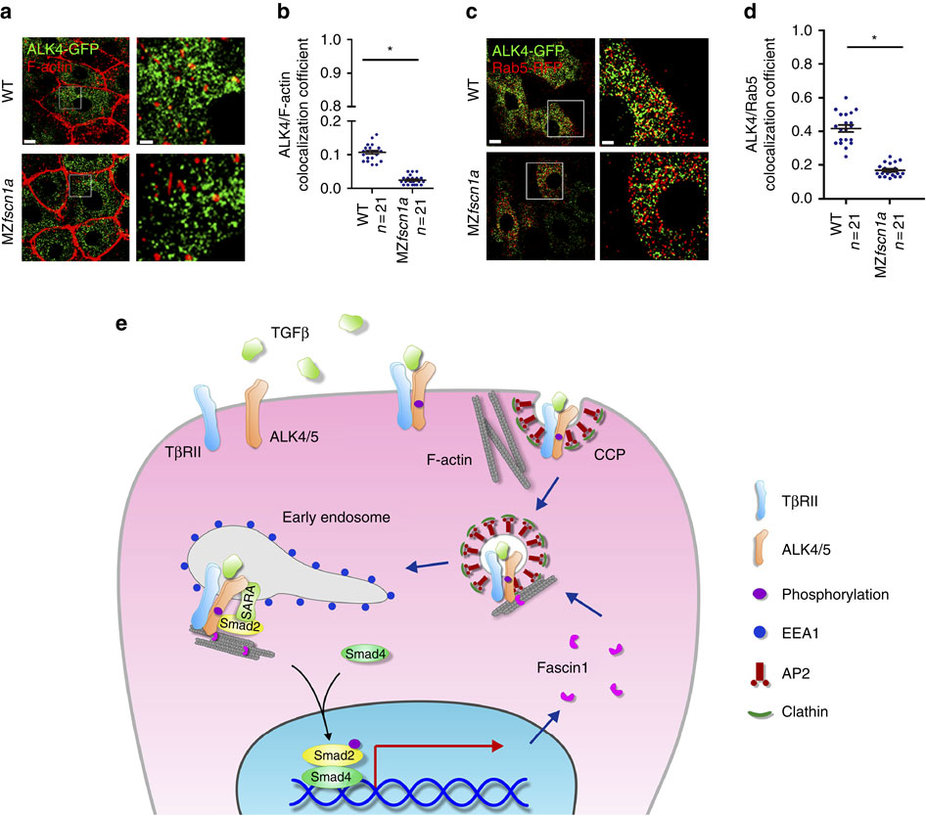
Fig. 8
The endocytic trafficking defects of Activin/Nodal type I receptor ALK4 in MZfscn1a mutants.
(a,b) 20 pg ALK4-GFP mRNA was injected into one blastomere of a 16-cell wild-type or MZfscn1a embryo. Embryos were harvested at shield stage and costained with phalloidin-TRITC and anti-GFP antibody to show the co-localization of F-actin (red) and ALK4 (green). GFP-positive hypoblast cells were selected and photoed using a Nikon A1R+ confocal microscope. The boxed area in the left image (Scale bar, 10 �m) is presented at a higher magnification in the corresponding right image (Scale bar, 2 �m) (a). Peason?s co-localization coefficient was quantified from the indicated cell numbers in three independent experiments and the group values are expressed as mean�s.d. Student?s t-test, *P<0.05 (b). (c) Immunostaining images of Rab5-RFP (red) and ALK4-GFP (green) in hypoblast cells of wild-type or MZfscn1a embryos. 20 pg ALK4-GFP mRNA and 20 pg Rab5-RFP mRNA were co-injected into one blastomere of a 16-cell wild-type or MZfscn1a embryo. Embryos were collected at shield stage for immunostaining with anti-GFP and anti-dsRed antibodies. Scale bar, left images, 10 �m; right images, 3 �m. (d) Peason?s co-localization coefficient of ALK4-GFP and Rab5-RFP was quantified from the indicated cell numbers in three independent experiments and the group values are expressed as mean�s.d. *P<0.05. (e) Proposed mechanism of the function of Fscn1 in trafficking and signalling of TGF-β type I receptors. TGF-β/Smad signal induces the expression of fscn1 gene and Fscn1 protein acting as a molecular linker between TGF-β family type I receptors and the actin cytoskeleton, which promotes the trafficking of internalized receptors from clathrin-coated vesicles to early endosomes, thereby enhancing TGF-β signal activity. TβRII, TGF-β type II receptor; CCP, clathrin-coated pit; AP2, assembly polypeptide 2.