Fig. S4
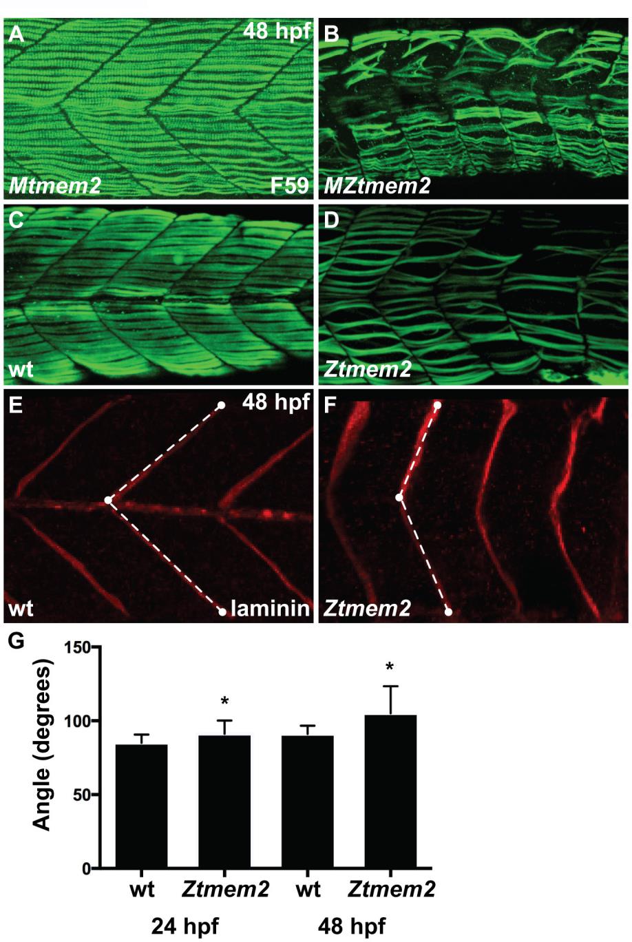
Impairment of muscle fiber organization in zygotic tmem2 mutants. (A-D) Immunofluorescence with F59 (green) reveals slow muscle fiber organization; lateral views with dorsal up at 48 hpf. At this stage, MZtmem2 mutants (B) exhibit detachment and disorganization of slow muscle fibers, in contrast to the normal attachment seen in their Mtmem2 siblings (A). Although we have not observed muscle fiber detachment in zygotic tmem2 (Ztmem2) mutants at 24 hpf (data not shown), we have found fiber detachment and disorganization in some Ztmem2 mutants (D) by 48 hpf (n=4 out of 33 Ztmem2 mutants examined). (E,F) Immunofluorescence detecting laminin (red) deposition at the MTJ reveals somite shape defects in Ztmem2 mutants. In wild-type (E) and Ztmem2 (F) sibling embryos, somite shape was evaluated by measuring the angle formed at the MTJ. White dots represent examples of the reference points chosen at the horizontal myoseptum, the dorsal edge of the MTJ, and the ventral edge of the MTJ; dashed lines represent the angle measured using ImageJ software. (G) Bar graph compares average angles formed at the MTJ in wild-type and Ztmem2 embryos at 24 hpf and 48 hpf (in wild-type, n=85 at 24 hpf and n=75 at 48 hpf; in Ztmem2, n=52 at 24 hpf and n=154 at 48 hpf). Error bars indicate s.d., and asterisks indicate a significant difference from wild-type (Student′s t-test; p<0.0001). Somite shapes in Ztmem2 mutants become less chevron-shaped and more U-shaped over time, presumably as maternal supplies of tmem2 are depleted and muscle fiber defects accumulate.

