
|
Fig. S4
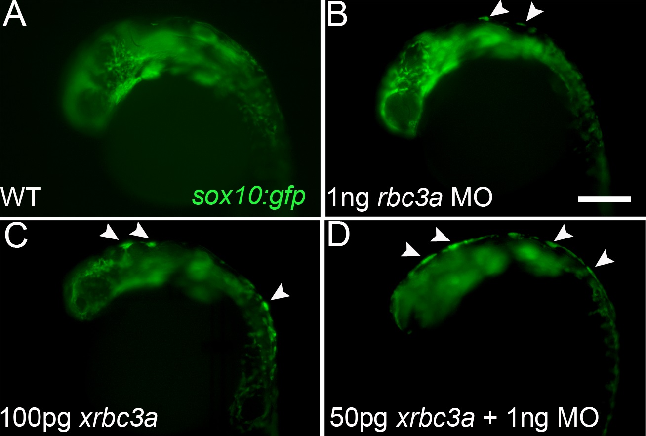
Injection of a 3′-truncated Xenopus rbc3a construct phenocopies Rbc3a loss of function. Fluorescent images of live sox10:gfp transgenics, lateral views, anterior to the left. (A, B) Injection of 1 ng/embryo of rbc3a-MO1 caused GFP+ cells to aggregate at the dorsal midline (arrowheads) by 24 hpf (41%, n = 7/17). (C) Injection of 100 pg/embryo of Xenopus rbc3a mRNA lacking 2.2 kb of the 3′ end of the ORF caused similar GFP+ dorsal aggregates (83%, n = 15/18). (D) Co-injection of 50 pg/embryo of truncated xrbc3a mRNA with 1 ng/embryo of rbc3a-MO1 increased the number and severity of embryos with GFP+ dorsal aggregates (79%, n = 11/14), with some embryos exhibiting a continuous strip of GFP+ cells all along the dorsal midline (36%, n = 5/14).