
|
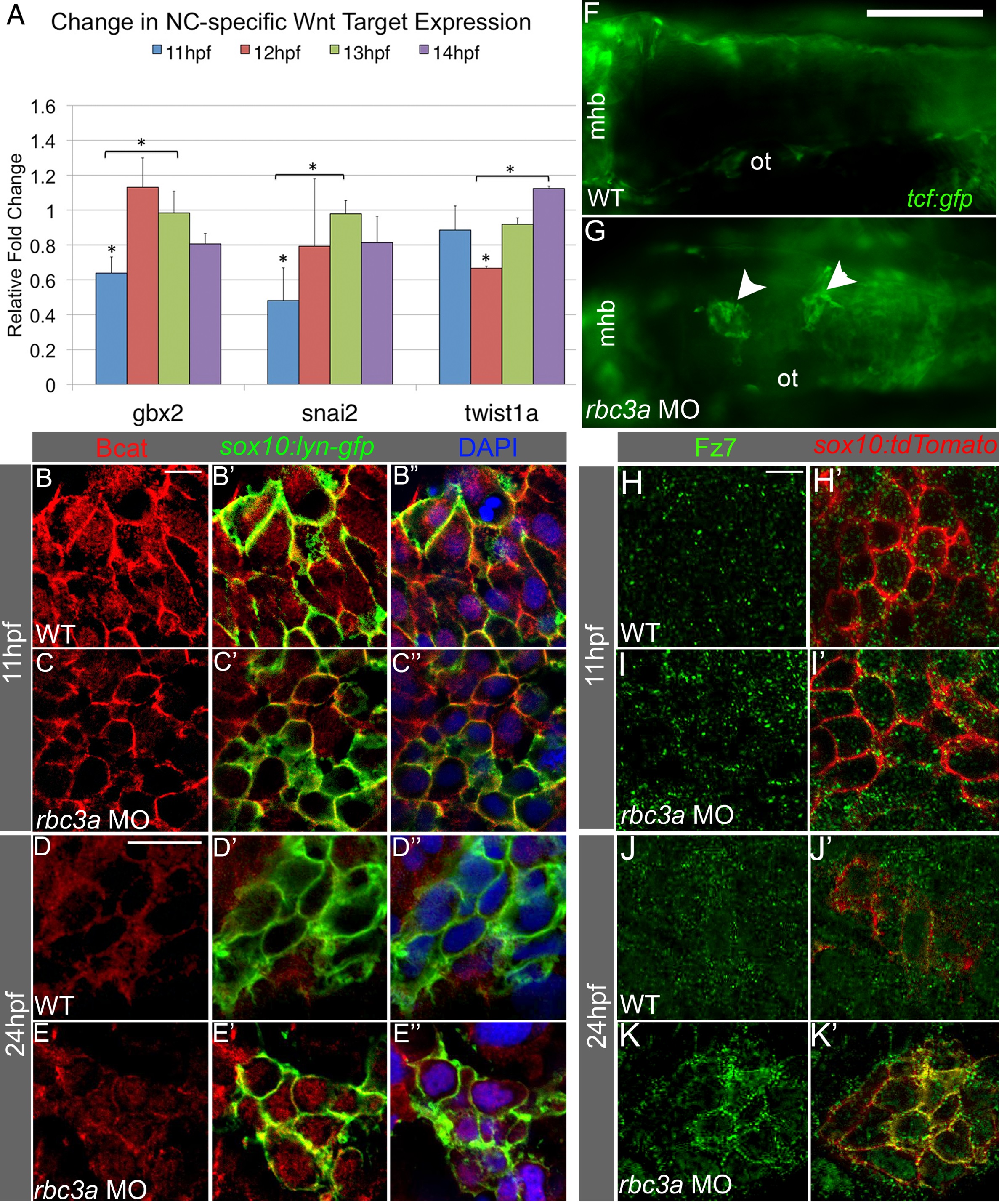
Fig. 5
Rbc3a knockdown disrupts subcellular localization of Bcat and Fz7 in NC cells.
(A) Quantitative RT-PCR analysis of early Wnt target genes with important roles in EMT reveals reduced expression of snai2, gbx2, and twist1a at 11-12 hpf in rbc3a-MO1-injected embryos. Error bars represent triplicate experiments � SEM. * p<0.05. (B-E3) Immunostaining with an anti-Bcat antibody (red) in whole-mounted, sox10:lyn-gfp transgenic embryos to label NC cell membranes (green) and DAPI to label nuclei (blue). Bcat levels in the nucleus are (B-C3) reduced in rbc3a-MO1-injected embryos at 11 hpf and (D-E3) elevated in the nucleus of MO-injected embryos at 24 hpf compared with wild-type (WT) controls. Scale bar, 10 �m. (F, G) Dorsal images of tcf:gfp Wnt reporter transgenic fish at 24 hpf identifies distinct aggregates of GFP+ cells (white arrowheads) in the dorsal midline of rbc3a-MO1-injected embryos (G) but not wild-type (F) embryos. Mhb, midbrain-hindbrain boundary; ot, otic vesicle. Scale bar, 100 �m. (H-K′) Immunohistochemical staining for GFP (green) after microinjection of fz7-yfp mRNA in sox10:lyn-tdtomato (red) transgenic embryos. At both 11 hpf (H-I′) and 24 hpf (J-K′), the number of YFP+ puncta per cell increased in NC cells in rbc3a-MO1-injected embryos. YFP colocalizes with tdTomato at the membranes of NC cells in rbc3a-MO1-injected embryos at 24 hpf (K′). Scale bar, 10 �m.