Fig. 1
Dynamics of Microglial Brain Colonization and Neuronal Apoptosis
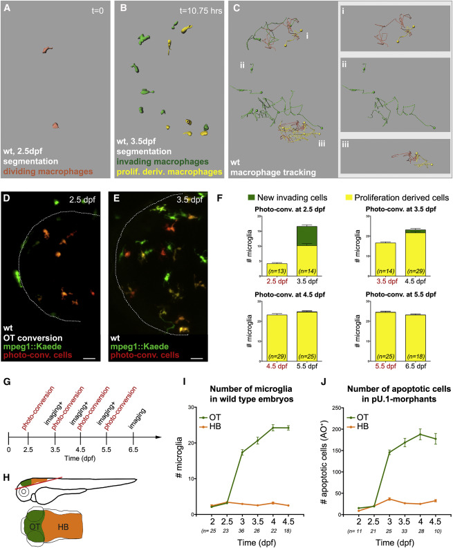
(A-C) Macrophage cell segmentation (A [t = 0 hr] and B [t = 10.75 hr]) and cell tracking (C) of a representative wild-type mpeg1::GAL4-UAS::Kaede embryo, imaged for 10.75 hr ( Movie S1). In orange are the proliferating and in green are the invading macrophages/tracks. Yellow marks cells resulting from division. Examples of single tracks are given in (i), (ii), and (iii).
(D and E) Dorsal view of a mpeg1::GAL4-UAS::Kaede embryo after microglia photo-conversion (D) and 24 hr later (E). Microglia that entered the brain after photo-conversion are green. Microglia derived from proliferating converted cells are in red/yellow. Dotted lines mark the tectal area.
(F) Quantifications of microglial photo-conversion at different developmental time points. In each graph, the histogram on the left represents the number of microglia photo-converted at a given time point (in red). The histogram on the right indicates the number of microglia entering the brain after conversion (green bar) or deriving from proliferation (yellow bar) 24 hr after photo-conversion.
(G) Experimental setup. Microglia are photo-converted at different time points. Then, 24 hr after each round of photo-conversion, green and red/yellow cells are quantified.
(H) Schematic illustrations of a 3-dpf embryo (side and dorsal views). The red line marks the depth at which confocal imaging was performed.
(I and J) Quantifications of the number of microglia (I) and apoptotic nuclei (J) in wild-type and pU.1-morphant brains, respectively, are shown.
OT, optic tectum; HB, hindbrain; AO, acridine orange; n, number of embryos. For all quantifications, data are pooled from three independent experiments. Scale bar represents 30 Ám. See also Figure S1 and Movie S1.

