
|
Fig. S4
LPC-G2A Pathway Is Required for Microglial Precursors Entering the Brain, related to Figure 4
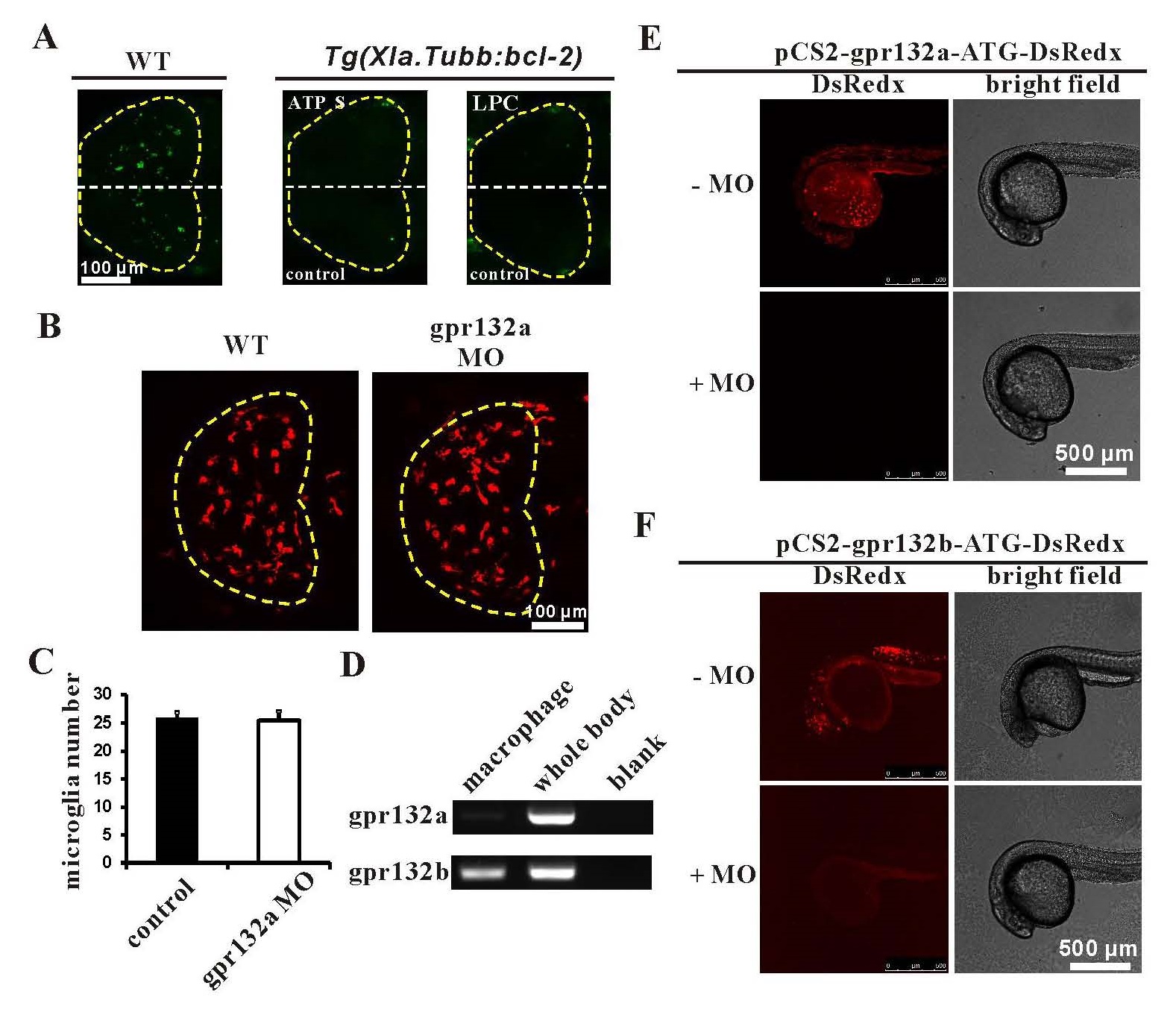
(A) AO staining showed that no excess apoptotic cells were observed in the brain injected with ATPγS or LPC, although the apoptotic cells could be readily observed in the brain of 3 dpf WT.
(B) Dorsal view images of the midbrain of Tg(mpeg1:loxP-DsRedx-loxP-eGFP) control and gpr132a morphants (MO). DsRedx+ cells represent microglia. Dashed lines indicate the midbrain.
(C) Quantification of the microglia number in the midbrain of Tg(mpeg1:loxP-DsRedx-loxPeGFP) control and gpr132a morphants (MO). n=8 for WT control and n=10 for MO. Error bars represent mean SEM.
(D) Examination of gpr132a and gpr132b expression in macrophage and whole fish body by RTPCR at 2.5-3 dpf.
(E) gpr132a MO efficiently blocked the expression of pCS2-gpr132a-ATG-DsRedx reporter construct.
(F) gpr132b MO efficiently blocked the expression of pCS2-gpr132b-ATG-DsRedx reporter construct.
Reprinted from Developmental Cell, 38(2), Xu, J., Wang, T., Wu, Y., Jin, W., Wen, Z., Microglia Colonization of Developing Zebrafish Midbrain Is Promoted by Apoptotic Neuron and Lysophosphatidylcholine, 214-22, Copyright (2016) with permission from Elsevier. Full text @ Dev. Cell