Fig. 1
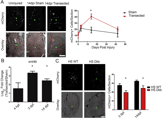
Wnt/β-catenin signaling is increased after spinal cord injury. (A) Representative images of Tg(7xTCFxla.siam:mCherryNLS) fish after sham surgery or spinal cord transection that were used for quantification of mCherry + cells. Graph shows the quantification of mCherry + nuclei at the indicated times. Green asterisk marks central canal. (B) Graph of the relative abundance of wnt4b transcripts as measured by using quantitative PCR from samples of spinal cords collected at the indicated times after spinal cord injury. (C) Representative images of Siam:mCherry x Hsp70l:Dkk double transgenic fish at 3 dpt. Graph shows the quantification of mCherry + nuclei times at indicated times and genotypes. Green asterisk marks central canal. *p < 0.05; scale bar, (A, C) 100 Ám; n ≥ 5 for (A), n = 3 for pooled samples in (B), n ≥ 3 for (C). Graphs (A, C) show mean ▒ SEM, graph (B) shows mean ▒ St Dev. (For interpretation of the references to colour in this figure legend, the reader is referred to the web version of this article.)

