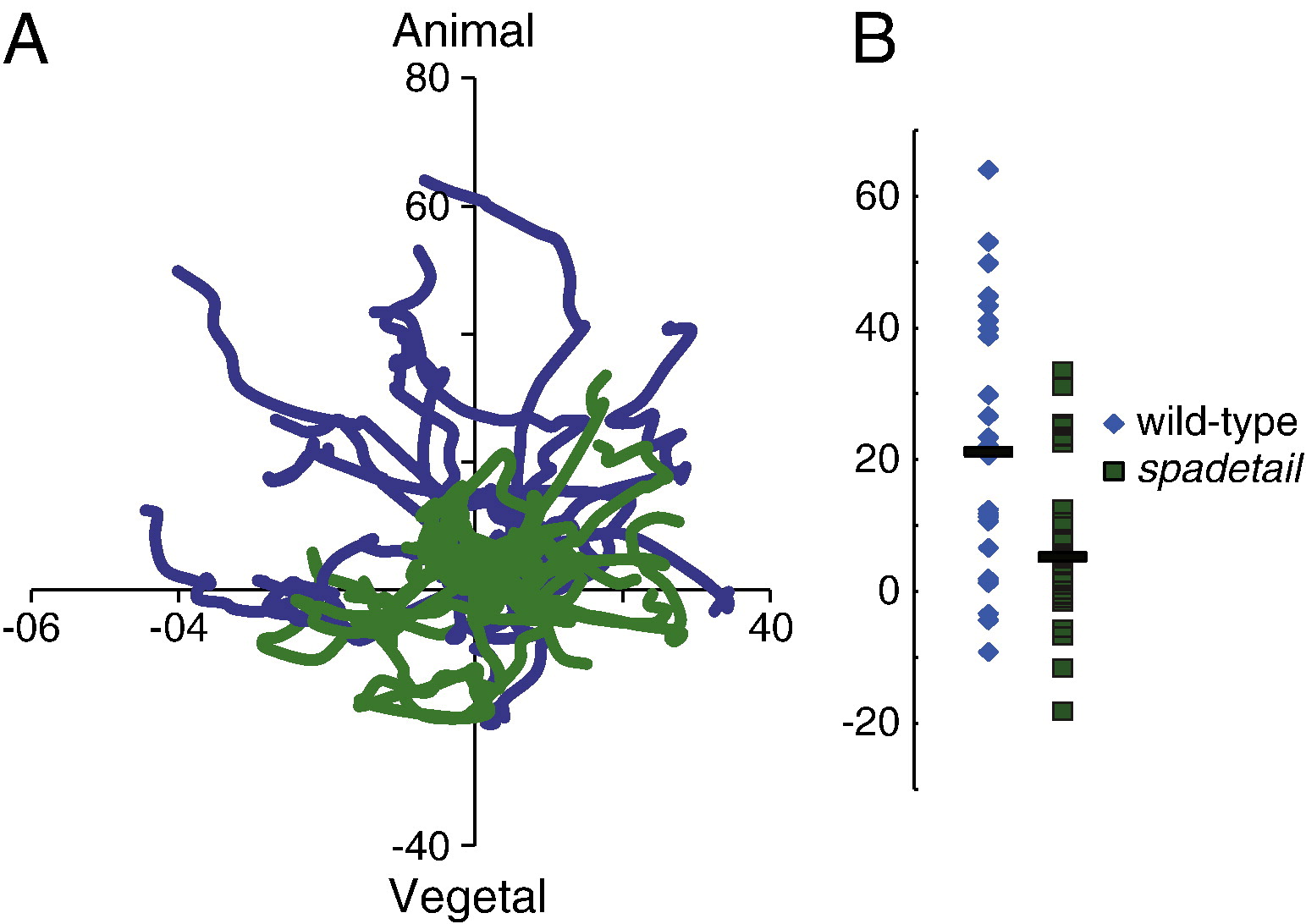
Fig. 3
spt morphant mesoderm migrates abnormally. (A) Recently involuted cells were tracked for 30 min in wild-type (blue traces) and spt morphant (green traces) embryos at 55-60% epiboly, lateral to the shield. The x-axis is aligned with the margin of the embryo. spt morphant cells failed to exhibit the animal-ward bias of migration observed in wild-type mesoderm. A representative set of tracks from one wild-type and one spt morphant is shown. (B) The final positions of all tracked cells on the y-axis are marked, with bars indicating median positions. Wild-type cells had a significant migration bias towards the animal pole, much greater than was observed in spt morphant cells. All axes are labeled with µm of displacement from the starting position. These results are taken from tracking 26 cells from 3 embryos, per condition.
Reprinted from Developmental Biology, 354(1), Row, R.H., Maître, J.L., Martin, B.L., Stockinger, P., Heisenberg, C.P., and Kimelman, D., Completion of the epithelial to mesenchymal transition in zebrafish mesoderm requires Spadetail, 102-110, Copyright (2011) with permission from Elsevier. Full text @ Dev. Biol.

