Fig. 5
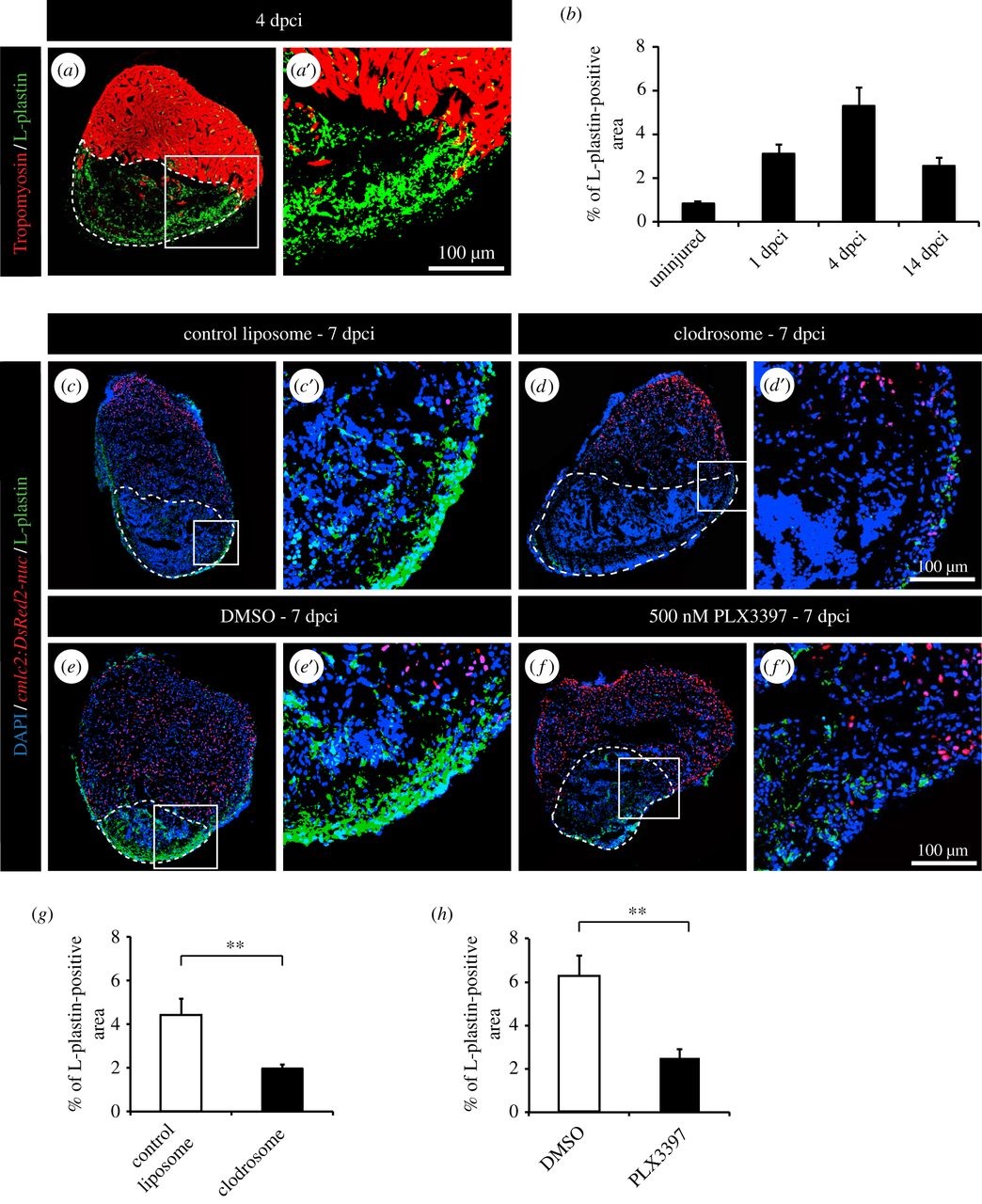
Clodronate liposome injections and PLX3397 treatment efficiently deplete phagocyte populations after cryoinjury. (a) Representative image of cryoinjured heart at 4 days post-cryoinjury (dpci) labelled with antibody against tropomyosin (red) to demarcate the remaining myocardium and L-plastin (green) to reveal leucocytes. The cryoinjured area is identified by the absence of tropomyosin (encircled by dashed line). (b) Quantification of the L-plastin-positive area in sections of uninjured hearts, and at 1, 4 and 14 dpci. (c-f) Representative images of hearts of transgenic fish cmlc2:DsRed2-nuc at 7 dpci with L-plastin staining (green). The cryoinjured area is identify by the absence of cmlc2:DsRed2-nuc expression (encircled by dashed line). (c,d) Hearts after control and clodronate liposome (clodrosome) injections. (e,f) Hearts after 0.05% DMSO or 500 nM PLX3397 treatment. (g,h) Quantification of L-plastin-positive area after clodronate liposome injection (g) and PLX3397 treatment (h) at 7 dpci. (n ≥ 4 hearts; ≥ 2 sections per heart; **p < 0.01).

