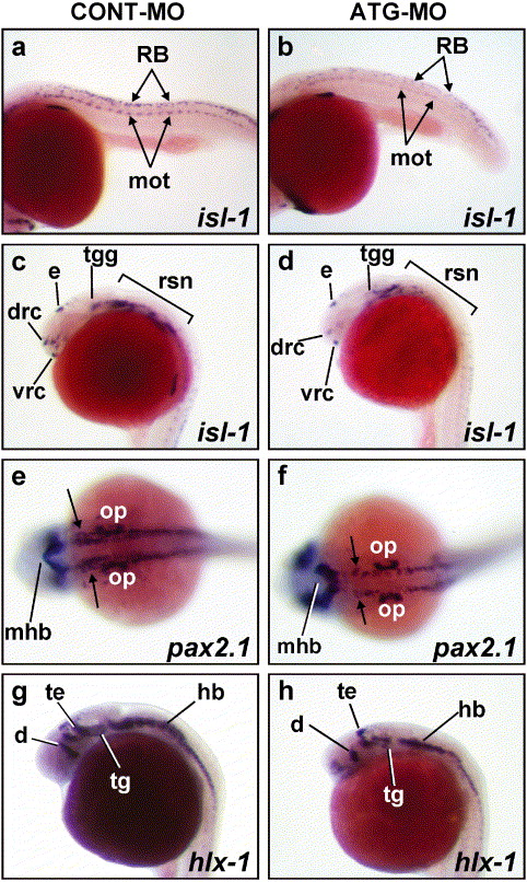
Fig. 5
Loss of differentiated neuronal populations in Neogenin morphants. (a-d) Compared to CONT-MO (3.7 ng)-injected embryos at 24 hpf (a), ATG-MO (3.7 ng)-injected embryos (b) displayed a reduction in isl-1 expression in primary motor neurons (mot) and Rohon-Beard sensory neurons (RB) along the entire length of the neural tube. In the brain primordia of morphants (d), isl-1-expressing neurons were also reduced in number including those in the dorsorostral cluster (drc) of the telencephalon, the trigeminal ganglia (tgg), and the reticulospinal neurons of the hindbrain (rsn) compared to control embryos (c). (f) A proportion of pax2.1-expressing neurons was absent from the hindbrain of ATG-MO-injected embryos (arrows) compared to control embryos (e). (g) In control embryos, hlx-1 was expressed by populations of neurons in the tegmentum (tg), tectum (te), diencephalon (d), hindbrain (hb), and neural tube. (h) Neogenin morphants exhibited a similar pattern of hlx-1-expressing cells in the dorsal region of the diencephalon (d), tectum, and tegmentum. (a-d, g, and h). Lateral views with rostral to the left. (e and f) Dorsal views with rostral to the left. mhb, midbrain-hindbrain boundary; op, otic placode.
Reprinted from Developmental Biology, 269(1), Mawdsley, D.J., Cooper, H.M., Hogan, B.M., Cody, S.H., Lieschke, G.J., and Heath, J.K., The Netrin receptor Neogenin is required for neural tube formation and somitogenesis in zebrafish, 302-315, Copyright (2004) with permission from Elsevier. Full text @ Dev. Biol.

