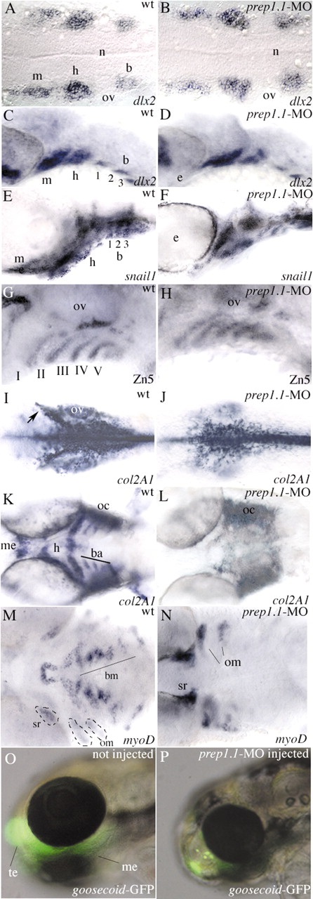
Fig. 7
Neural crest cells migrate to the pharyngeal arches in prep1.1 morphants but do not differentiate into chondrocytes. (A,B) At 19 hpf (20-somite stage), dlx2 expression reveals three discrete clusters of neural crest cells migrating to the mandibular (m), hyoid (h) and branchial (b) arches in wild-type embryos and prep1.1 morphants. (C,D) At 33 hpf, in both wild-type and prep1.1-MO-injected embryos the dlx2-positive neural crest cells have reached the target position; the branchial cluster has split into three subgroups of cells. (E,F) At 48 hpf, snail1-expressing cells have segregated in the segmented pharyngeal arches both in wild-type embryos and prep1.1 morphants. (G,H) At 48 hpf the pharyngeal endoderm, revealed by the Zn5 antibody, is correctly formed in prep1.1 morphants. (I,J) At 28 hpf, col2a1 expression in prep1.1-MO injected embryos reveals normal differentiation of neurocranium cartilages. However, the mesenchyme originating the parachordalia of the neurocranium is reduced (cells indicated by an arrow in I). (K,L) At 48 hpf, in the pharyngeal region of prep1.1 morphants col2a1 is expressed only in the otic capsule; expression is lacking in branchial arches. (M,N) At 3 dpf, in the pharyngeal region of prep1.1 morphants myoD is expressed only in opercular muscles and superior rectus of the eye; expression is lacking in branchial muscles. (O,P) prep1.1 knockdown in gsc::GFP transgenic larvae suppresses GFP expression in the first pharyngeal arch but not in the anterior telencephalic area. Embryos are in dorsal (A,B,I-N) and lateral (C-H,O,P) views, with anterior to the left. b, branchial arch; ba, branchial arches; bm, branchial muscles; h, hyoid arch; m, mandibular arch; me, Meckel′s cartilage; n, notochord oc, otic capsule; om, opercular muscles; ov, otic vescicle; sr, superior rectum of the eye; te, telencephalon. Quantitative data: (picture) probe, defective/total; (B) dlx2, 1/18; (D) dlx2, 0/22; (F) snail1, 4/25; (H) zn5, 4/18; (J) col2a1, 18/21; (L) col2a1, 18/19; (N) myoD, 16/18; (P) gsc, 4/5.

