Fig. 4
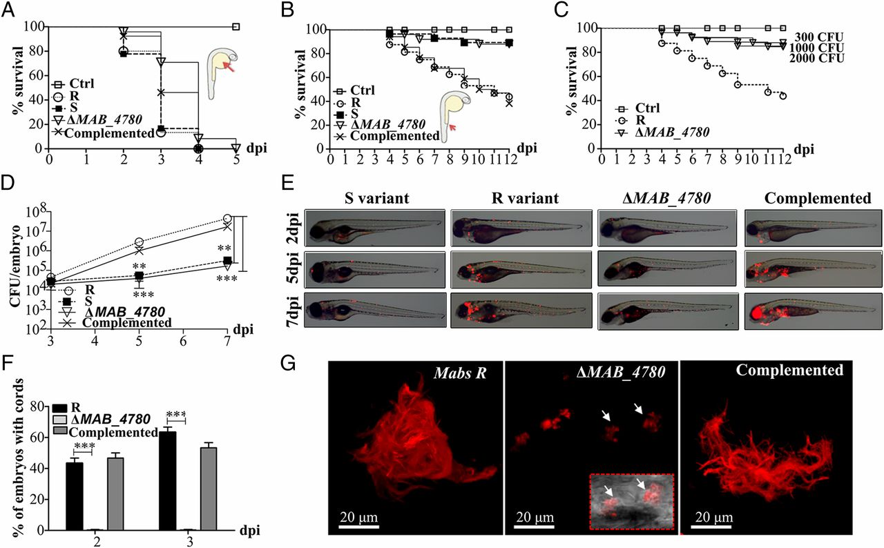
The ΔMAB_4780 mutant is extremely attenuated in infected zebrafish embryos. (A) Survival curves of embryos injected in the yolk sac (~100-200 cfu) of tdTomato-expressing Mabs S, Mabs R, ΔMAB_4780 mutant, or the complemented strain (n = 20). There are no significant differences between the different infected strains. Data are representative of three independent experiments. The Inset represents an embryo at 30 hpf, and the arrow indicates the injection site in the yolk. Ctrl, noninfected control embryos. (B) Survival curve for zebrafish embryos (n = 20) injected i.v. at 30 hpf with 100-200 cfu of the different Mabs strains compared with control embryos. Larvae infected with the ΔMAB_4780 mutant showed a significant increase in survival compared with larvae infected with the parental strain (P = 0.0008, log-rank test), whereas the complemented strain behaved like the wild-type R variant and restored virulence (no statistically significant differences were seen in the survival of embryos infected with the R or complemented strains; log-rank test). Shown are representative data of three independent experiments. The Inset represents an embryo at 30 hpf, and the arrow indicates the caudal vein injection site. (C) Survival curve of embryos infected with increasing doses of the ΔMAB_4780 mutant. Embryos injected with the ΔMAB_4780 mutant showed no statistically significant difference with the control group in terms of survival, regardless of the dose. Data shown are representative of two independent experiments (n = 20). (D) In vivo growth of the ΔMAB_4780 mutant. Enumeration was performed by plating homogenates of whole individual larvae at different time points on selective agar plates and subsequent counting of bacterial colonies. A significant reduction in bacterial burden was observed with the ΔMAB_4780 mutant (Kruskal-Wallis with Dunn?s multiple test; **P < 0.01; ***P < 0.001). Data are representative of three independent experiments. (E) Bright field/fluorescence overlay images of tdTomato-labeled Mabs (<150 cfu). Each larva was followed and imaged at 2, 5, and 7 dpi. (F) Cords were recorded in i.v.-infected embryos with either tdTomato-expressing Mabs R, ΔMAB_4780 mutant, or complemented strains at 2 and 3 dpi. Cords were never observed in ΔMAB_4780-infected embryos. Histograms represent means calculated from three independent experiments (n = 10 per experiment), and the statistical test used was Fisher?s exact test; ***P < 0.001. Error bars represent the SEM. (G) Maximum intensity projection of confocal images showing representative pathological events in 3 dpi embryos i.v.-infected with Mabs R, ΔMAB_4780, or complemented strains expressing tdTomato (100-200 cfu). Arrows indicate infected cells containing the tdTomato-labeled ΔMAB_4780 mutant. Only Mabs R and complemented strains formed cords.

