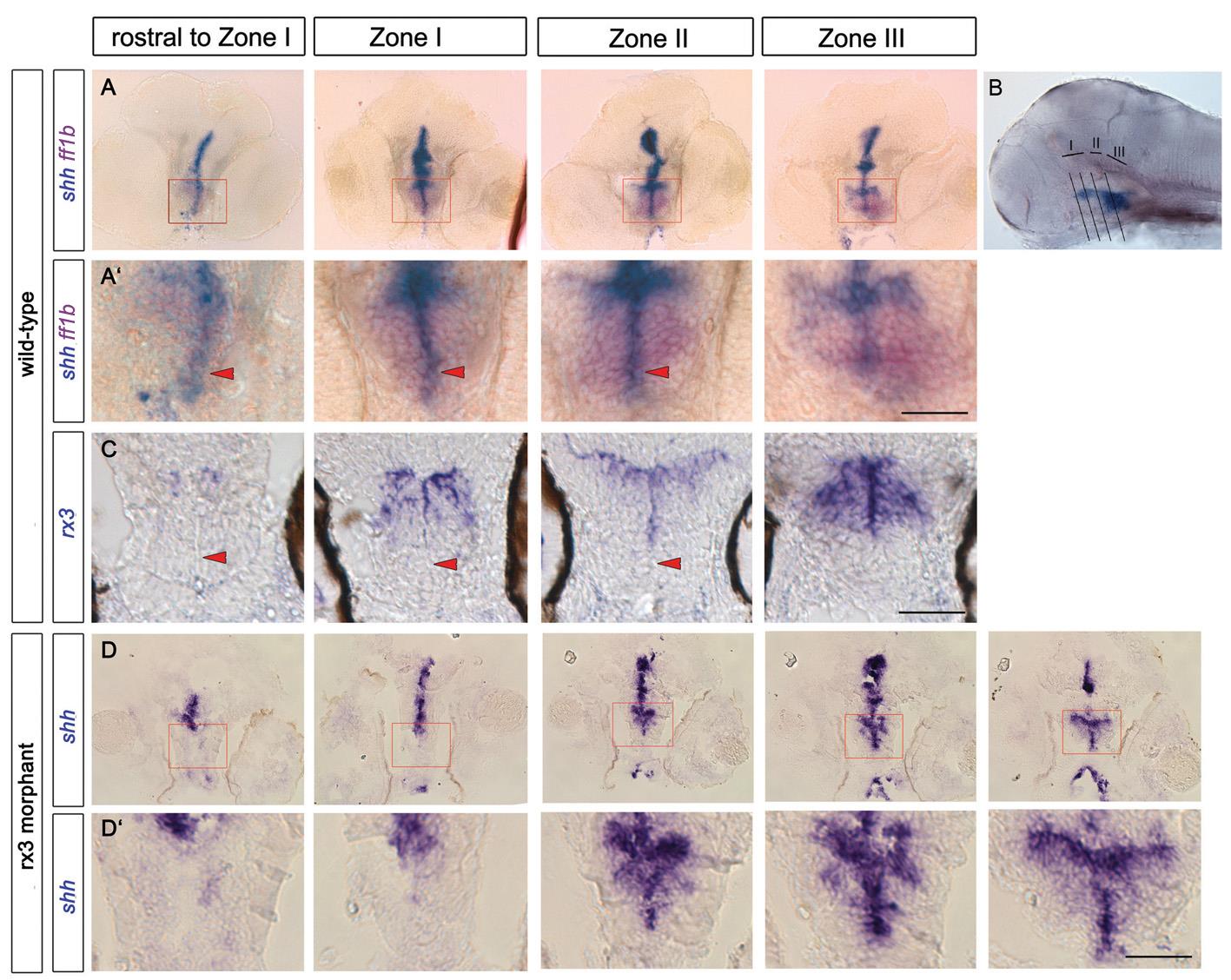
Fig. S1
Loss of AR and disorganization of lateral recess in rx3 morphant. (A-C) Transverse sections (A,C) through 55hpf control embryos, at planes shown in (B) after in situ hybridization with shh/sf1 (A) or rx3 (C). (A′) shows high-power views of boxed regions in (A). AR cells are shh+rx3- (red arrowheads in A′,C). (D) Transverse sections at equivalent positions in rx3 morphant embryos: the shh+ AR fails to form (D,D′, left hand panels), shh+ progenitors accumulate abnormally in/around the 3rd ventricle (middle panels). In more posterior regions of the neuraxis, shh appears normal (right hand panel). Scale bars: 50Ám.

