
|
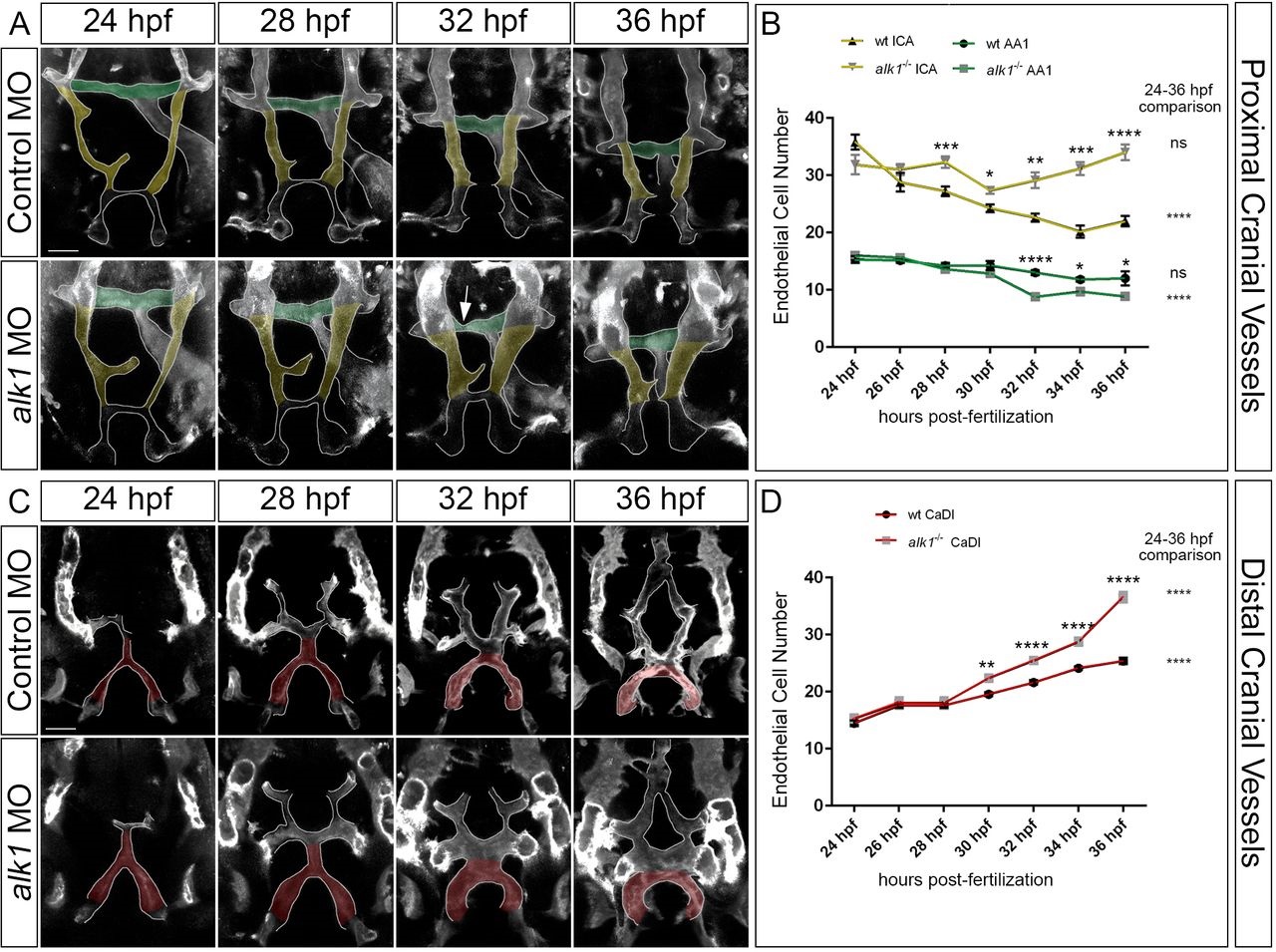
Fig. 2
Arterial endothelial cell numbers are altered in alk1-deficient embryos in a segment-specific manner. (A,C) 2D maximum projections of selected time points from confocal time-lapse imaging of Tg(fli1a.ep:mRFP-CAAX)pt505 control and alk1-morphant embryos, 24-36hpf, showing proximal cranial vessels/ventral planes (A) and distal cranial vessels/dorsal planes (C) of a single embryo. Dorsofrontal views, anterior down. Shaded regions correspond to vessel segments analyzed for cell number in B,D: green, AA1; yellow, proximal ICA; red, CaDI. Arrow in A points to transient AA1 stenosis. Scale bars: 50�m. (B,D) Endothelial cell counts in AA1 and ICA (B) and CaDI (D) in precisely staged alk1-/- embryos and wild-type (wt) siblings. Data are mean�s.e.m., ne4 embryos for each data point. Data were analyzed by two-way ANOVA followed by Bonferroni′s multiple comparisons test. Wild type and alk1-/- comparisons: significance indicated above time point. Within-treatment temporal comparisons: significance indicated to right of graph. ns, not significant, *P<0.05, **P<0.01, ***P<0.001, ****P<0.0001.