
Image
|
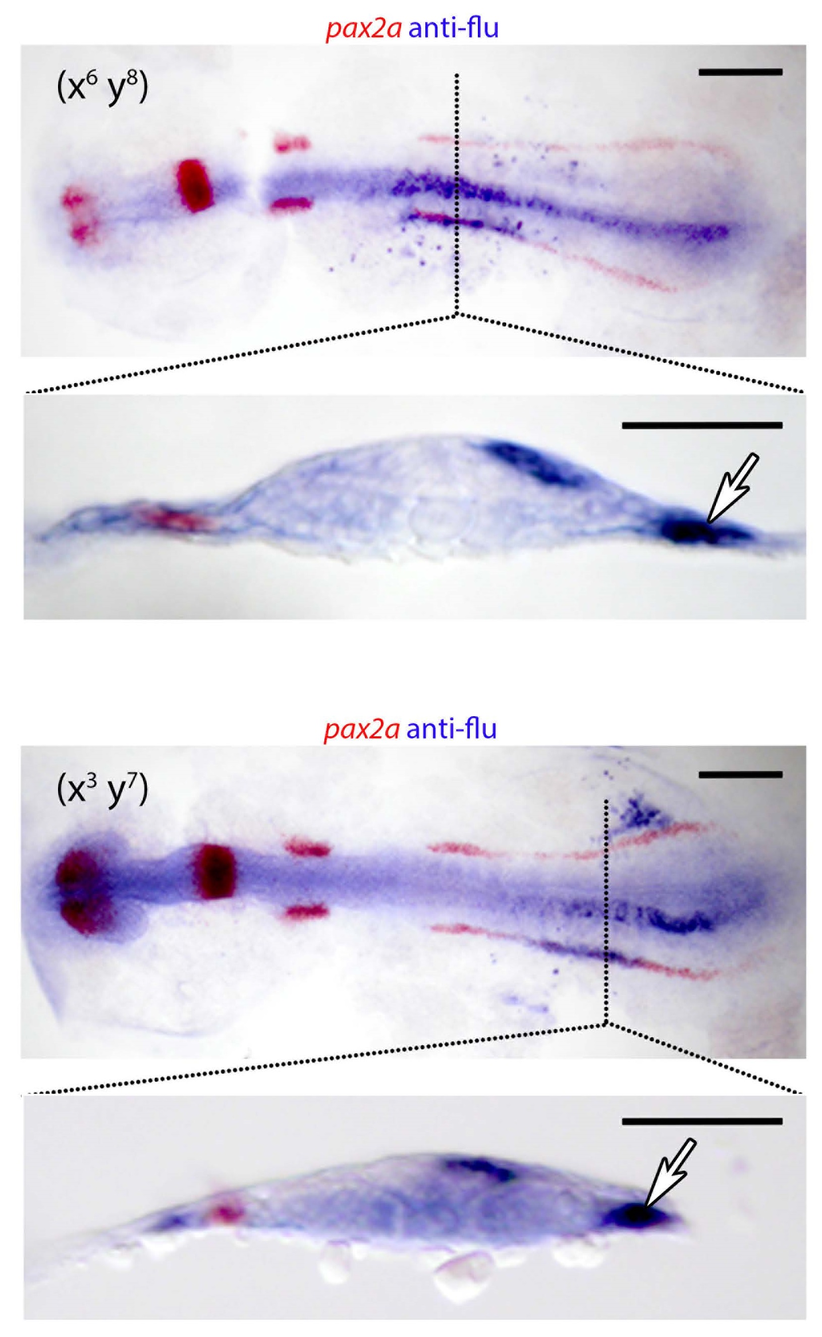
Figure Caption
Fig. S5
Cross-sections of fate-mapped embryos double-stained for pax2a transcripts.
Anterior and posterior nephron progenitors were lineage labeled in 85% epiboly embryos, examined by whole mount in situ hybridization at the 10 somite stage, and sectioned. Labeled cells contributing to the proximal and distal nephron were identified using an antibody to uncaged fluorescein (purple) and antisense probe for pax2a (red). Flat-mounted embryos are shown with anterior to the left, transverse sections are orientated with uncaged side to the right. Scale bars represent 200µm
Acknowledgments
This image is the copyrighted work of the attributed author or publisher, and
ZFIN has permission only to display this image to its users.
Additional permissions should be obtained from the applicable author or publisher of the image.
Full text @ Nat. Commun.