
|
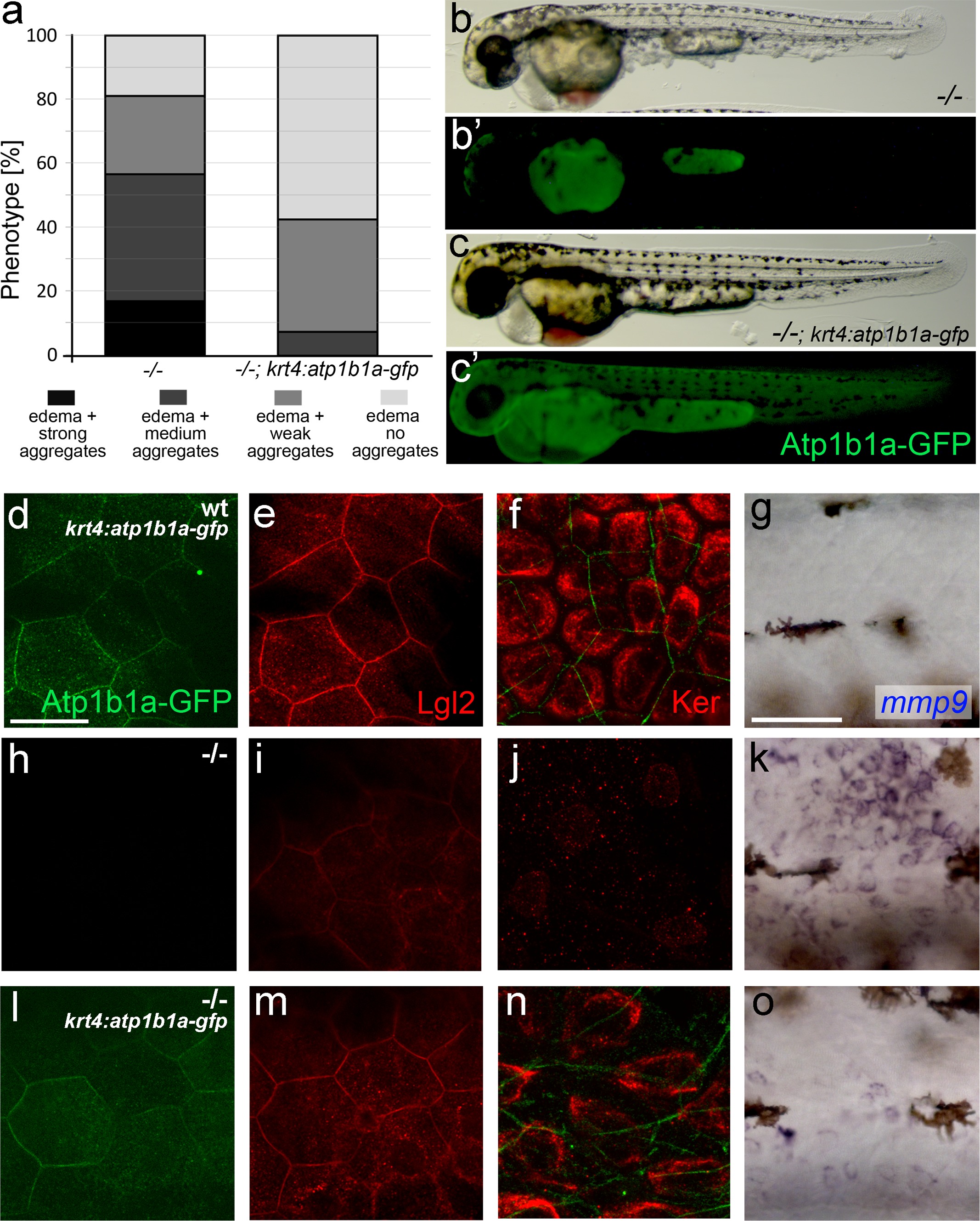
Fig. 8 S1
Tg(krt4:atp1b1a-gfp)-driven atp1b1a expression in the periderm of psoriasis mutants kept in hypotonic medium leads to a partial rescue of the polarity defects and malignant transformation in basal keratinocytes.
(a) Quantification of phenotypes of non-transgenic psoriasis mutants and psoriasis mutants carrying the krt4:atp1b1a-gfp transgene, raised in hypotonic E3, at 54 hpf. For classification of phenotypic strengths, see Figure 9a-d. (b-c) Representative live images of a psoriasis mutant lacking the transgene (no peridermal GFP; b′), which has pericardial edema and epidermal aggregates (b), and a psoriasis mutant expressing the transgene (peridermal GFP; c′), which has pericardial edema but no epidermal aggregates (c). (d-o) IF of GFP (transgene-encoded Atp1b1a-GFP) in periderm (green; d,h,l; 54 hpf), Lgl2 in periderm (red; e,i,m; 54 hpf) and cytokeratin in basal keratinocytes (red; f,j,n; 84 hpf), and WISH of mmp9 transcripts in basal keratinocytes (g,k,o; 54 hpf) of transgenic wt siblings (d-g), non-transgenic psoriasis mutants (h-k) and psoriasis mutants carrying the krt4:atp1b1a-gfp transgene (l-o). Periderm-specific expression of atp1b1a in mutant embryos leads to a partial restoration of the polarity markers Lgl2 and cytokeratin, and to a reduction of the malignancy marker mmp9. However, obtained levels are still higher than in the wt sibling control. For the reduction of the malignancy marker pAKT, see Figure 8f-h. Scale bars: 20 �m (d-f, h-j. l-n) and 50 �m (g, k, o).