Fig. 6
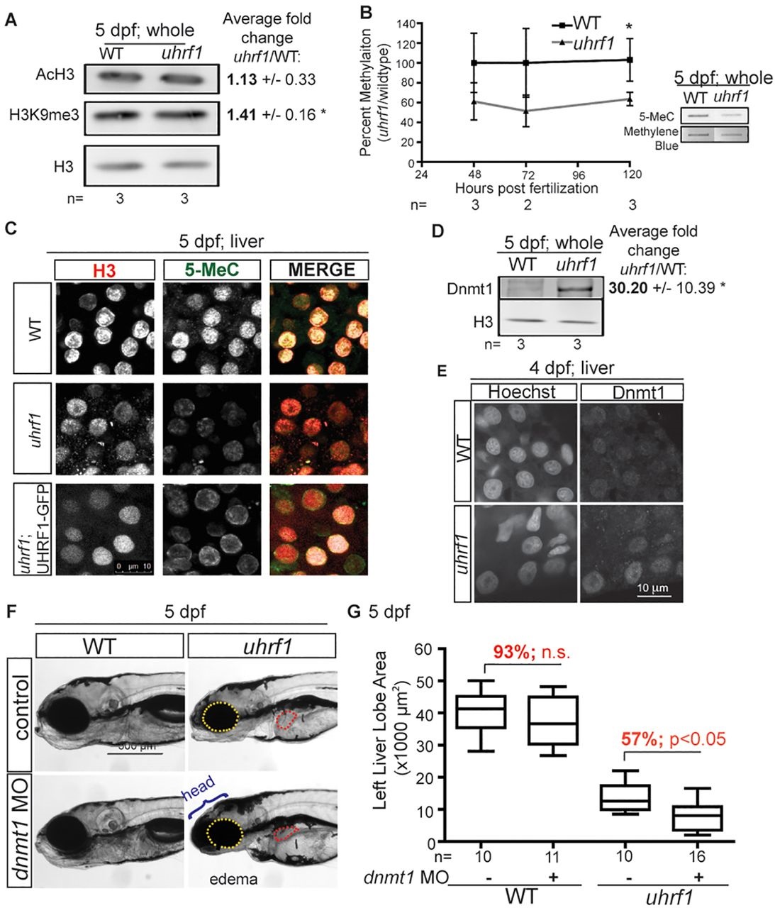
The uhrf1 mutant phenotype is mediated by hypomethylation. (A) Western blot of AcH3 and H3K9me3 levels in whole 5dpf larvae normalized to H3. Average ratios to H3 from three clutches are indicated with the s.d. *P<0.05 by Student′s t-test. (B) 5-MeC levels in uhrf1 mutant embryos and wild-type siblings, as measured by slot blot from at least three clutches. Mutant embryos were identified by genotyping at 48-72hpf and sorted based on morphological criteria at 96-120hpf. (C) 5-MeC immunofluorescence on livers of 5dpf wild type, uhrf1 mutants and Tg(fabp10:UHRF1-EGFP)Low;uhrf1 mutants. (D) Western blot of Dnmt1 in whole embryos. Average ratio of Dnmt1 to H3 in three clutches is indicated with the s.d. *P<0.05 by Student′s t-test. (E) Dnmt1 immunofluorescence and Hoechst staining for DNA in livers of 4dpf larvae. (F) Enhancement of the phenotypes in uhrf1 mutants injected with dnmt1 morpholino are indicated. (G) Quantification of left liver lobe size in 10-17 larvae on 5dpf. Top and bottom of box plot designate 75% and 25% of the population, respectively, separated by the median; bars represent 10th and 90th percentiles. *P<0.05. The percentage of the average liver size in dnmt1 morphants compared with the control injected larvae is indicated in red. n.s., not significant.

