
Image
|
Figure Caption
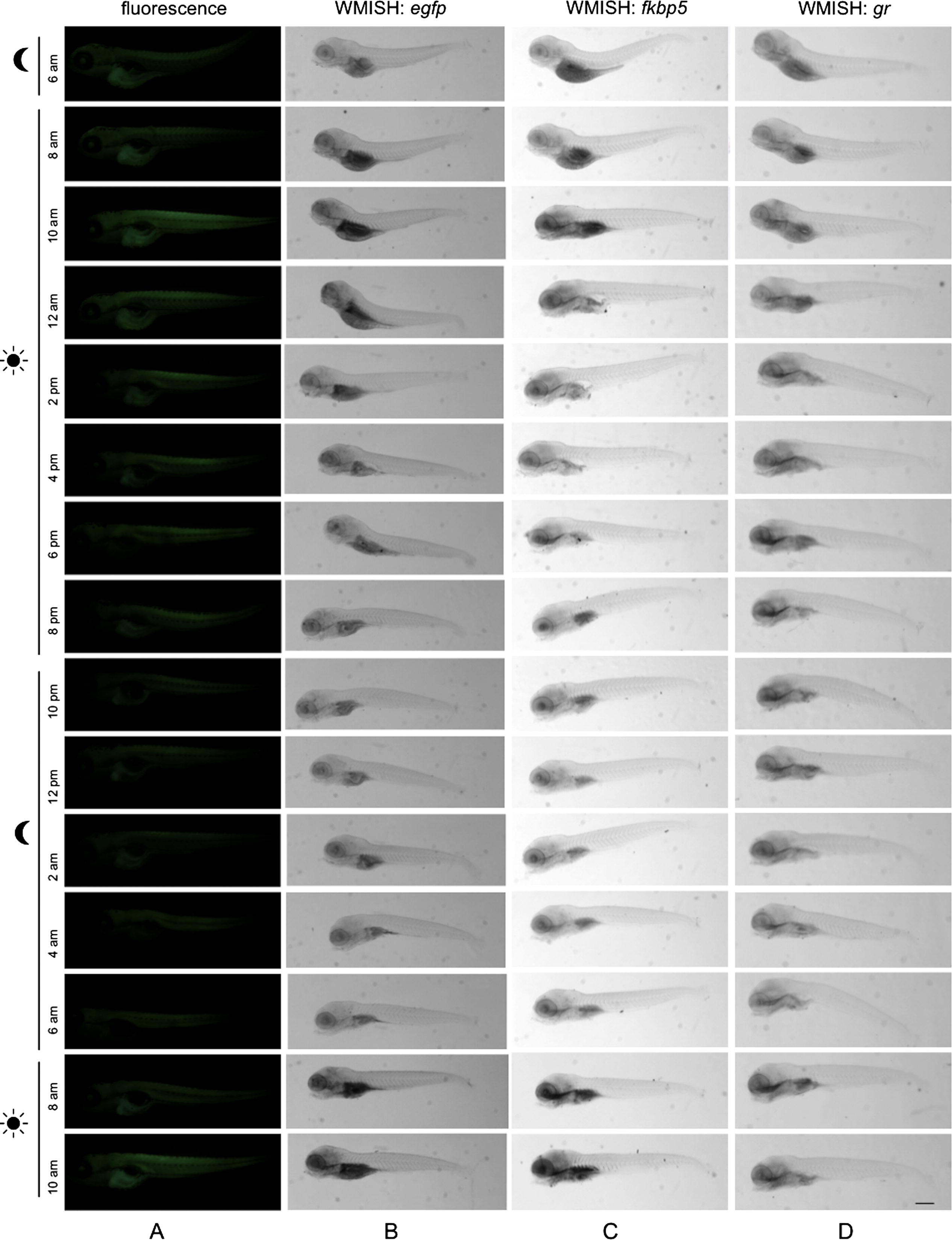
Fig. 8
(A) Fluorescence microscopy lateral view, (B) WMISH of egfp mRNA of 5-dpf transgenic larvae and (C) WMISH of fkpb5 mRNA, (D) WMISH of gr mRNA of 5-dpf WT larvae exposed to standard photoperiodic regime and analyzed from 2 h before light onset for 28 h. Scale bar: 200 �M.
Figure Data
Acknowledgments
This image is the copyrighted work of the attributed author or publisher, and
ZFIN has permission only to display this image to its users.
Additional permissions should be obtained from the applicable author or publisher of the image.
Reprinted from Molecular and Cellular Endocrinology, 392(1-2), Benato, F., Colletti, E., Skobo, T., Moro, E., Colombo, L., Argenton, F., Dalla Valle, L., A living biosensor model to dynamically trace glucocorticoid transcriptional activity during development and adult life in zebrafish, 60-72, Copyright (2014) with permission from Elsevier. Full text @ Mol. Cell. Endocrinol.