Fig. 5
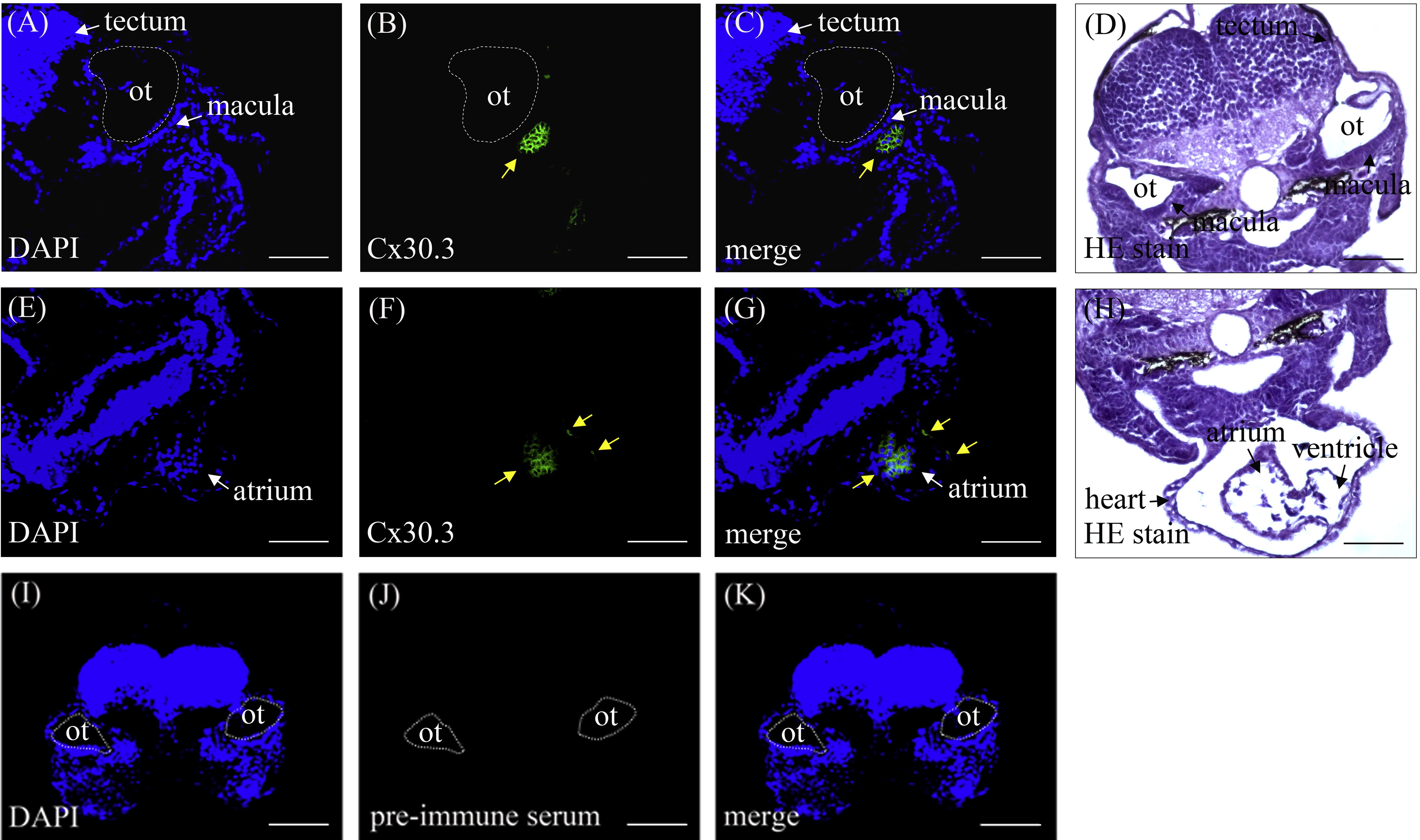
Zebrafish Cx30.3 proteins expressed in the heart and inner ear of 72 hpf embryos. The sections from paraffin-embedded 72 hpf embryos were immunodetected with antibodies against Cx30.3 and counterstained using HE staining. Zebrafish Cx30.3 proteins, indicated by yellow arrows, were shown in the macular cells of inner ear (A-D) and in the heart (E-H) using anti-Cx30.3 antibody. However, Cx30.3 proteins were not detected by pre-immune serum (I-K). (A, E, I) Sections from the 72 hpf embryos were counterstained with DAPI to highlight the nuclei. (B, F, J) Cx30.3 proteins expression. (C, G, K) Panel A, E or I merges with panel B, F or J respectively. (D, H) HE staining. ot: otic vesicle. Scale bars represent 50 Ám. (For interpretation of the references to color in this figure legend, the reader is referred to the web version of this article.)
Reprinted from Hearing Research, 313, Chang-Chien, J., Yen, Y.C., Chien, K.H., Li, S.Y., Hsu, T.C., Yang, J.J., The connexin 30.3 of zebrafish homologue of human connexin 26 may play similar role in the inner ear, 55-66, Copyright (2014) with permission from Elsevier. Full text @ Hear. Res.

