Fig. S3
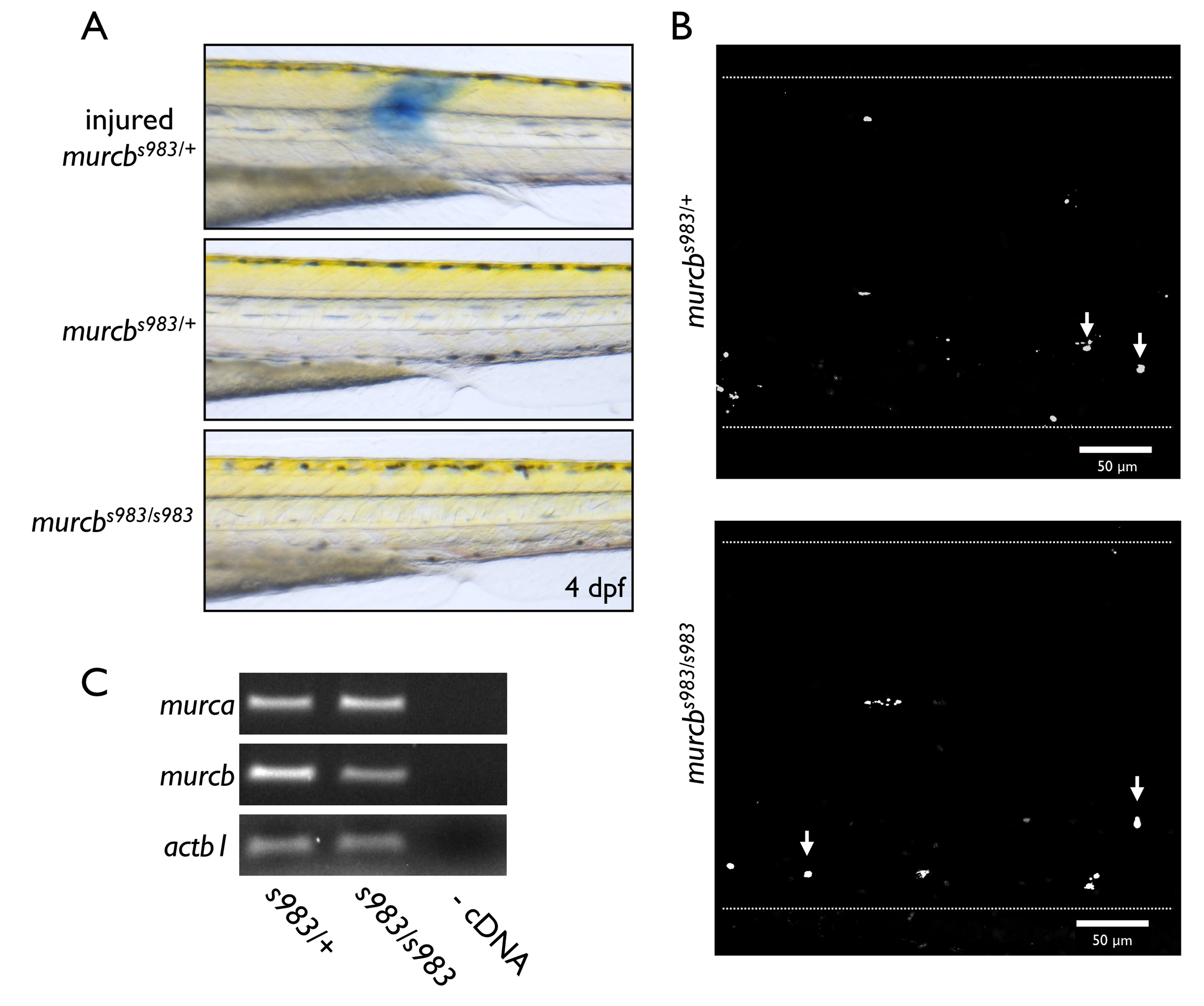
Analysis of Cavin4b/Murcb deficient zebrafish.
A. Evans blue dye assay of membrane integrity. The top panel is an injured somite and is shown as a positive control. No significant changes were observed between mutants and heterozygous controls. B. Representative maximal projection confocal images from live whole mount acridine orange staining of murcbs983/+ and murcbs983/s983 zebrafish trunk at 80 hpf. Arrows point to DNA fragmentation. Dotted lines outline the larvae. No significant changes were observed between mutants and heterozygous controls. C. RT-PCR analysis of murca and murcb mRNA from murcbs983/+ and murcbs983/s983 larvae at 72 hpf. No significant changes were observed between mutants and heterozygous controls.

