
|
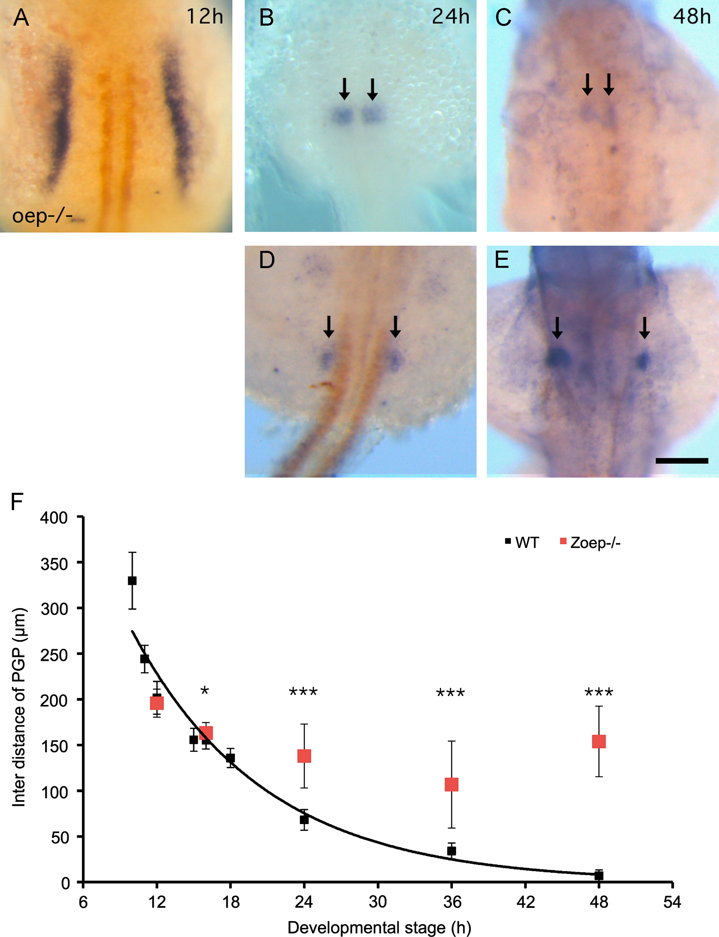
Fig. 3
Varied PGP phenotypes in Zoep-/- embryos. (A) Wt1a (blue) and myoD (orange) double in situ shows normal PG pattern at 12 hpf (6-somite) in Zoep-/-embryos. Both normal (B) and abnormal (D) PG (arrows) phenotype is observed at 24 hpf. Mild (C) and severe PG (arrows) midline convergence defect is observed at 48 hpf. All images are dorsal view anterior to the top and at the same magnification. The scale bar in E indicates 100 �m. (F) Zoep-/- inter distance of PGP (red squres) is compare to WT on it2s plot at 12 (n=40, P=0.15), 16 (n=24), 24 (n=22), 36 (n=34), 48 (n=29) hpf. P<0.05, *P<0.0005, ***Error bars represent SD.
Reprinted from Developmental Biology, 384(2), Huang, C.J., Wilson, V., Pennings, S., MacRae, C.A., and Mullins, J., Sequential effects of spadetail, one-eyed pinhead and no tail on midline convergence of nephric primordia during zebrafish embryogenesis, 290-300, Copyright (2013) with permission from Elsevier. Full text @ Dev. Biol.