
|
Fig. 5
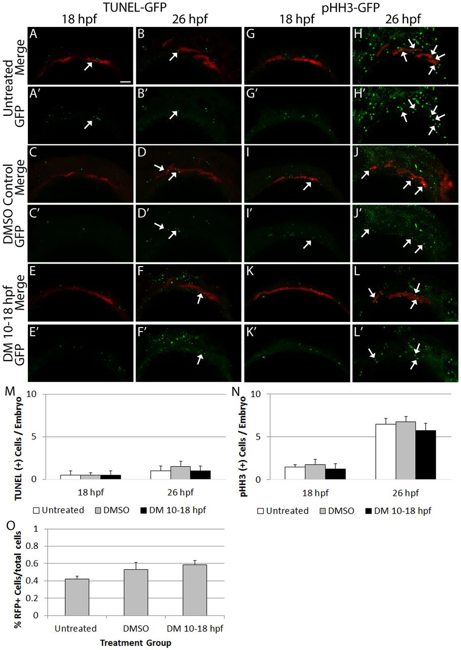
Blocking Bmp signaling does not alter endodermal cell number. (A-F′,M) Confocal images of sox17:dsred transgenic embryos labeled for cell death at 18 and 26hpf. Similar, low levels of apoptosis are present across all groups (18hpf, f=0, P=1; 26hpf, f=0.3, P=0.6, one-way ANOVA, n=4 embryos). (G-L′,N) Confocal images of sox17:dsred transgenic embryos labeled for cell proliferation at 18 and 26hpf. No differences in the level of proliferating endoderm cells are apparent (18hpf, f=0.2, P=0.8; 26hpf, f=0.1, P=0.8, one-way ANOVA, n=4 embryos). Arrows indicate overlap between the transgene and marker expression. Lateral views, anterior to the left. (O) Total endoderm cell numbers were counted via FACS and no statistically significant differences between 26hpf Dorsomorphin-treated, untreated or DMSO-treated embryos were observed (f=1.8, P=0.2, one-way ANOVA, n=8 samples, 10 embryos per sample). Error bars indicate mean+s.e.m. Scale bar: 50�m.