
|
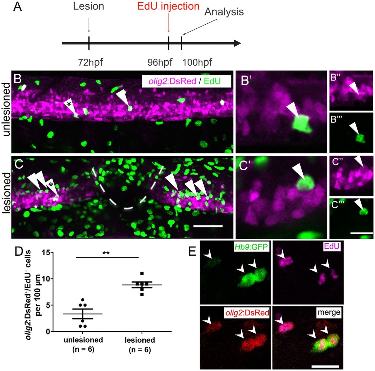
Fig. 2
After a lesion, the pMN domain shows increased proliferation and gives rise to motor neurons. (A) Time line of the experiment. (B,C) olig2:DsRed+ cells (arrowheads) in the pMN domain that incorporated EdU within the last 4h. (B′-C′′′) Higher magnifications of single optical sections of the cells indicated by asterisks in B and C, respectively, showing double labelling. (D) The number of proliferating cells in the pMN domain is significantly increased in the vicinity of the lesion site (Mann-Whitney test; **P=0.0049). (E) In Hb9:GFP and olig2:DsRed double-transgenic larvae (lesion: 3dpf; analysis: 5dpf), newly generated motor neurons (Hb9:GFP+/EdU+) that retain DsRed protein are indicated by arrowheads. Lateral views are shown; rostral is left, dorsal is up. Values are means±s.e.m. Scale bars: 50µm in C for B,C; 20µm in C′′′ for B′′-C′′′ and 10µm for B′,C&prime& 15µm in E.