
|
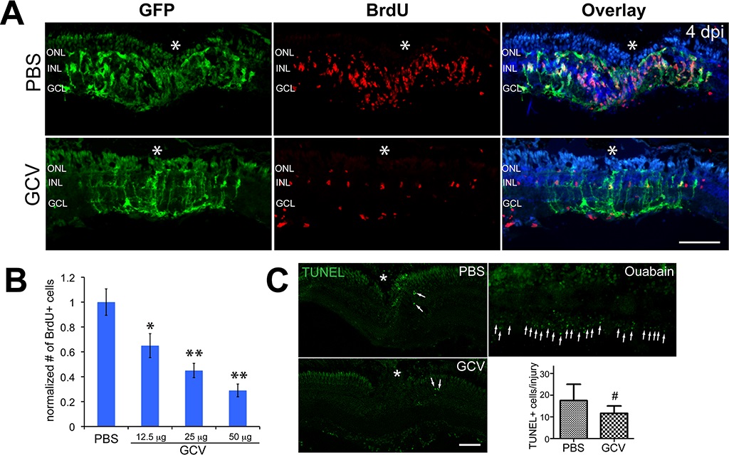
Fig. 1
Ganciclovir treatment significantly reduced the number of MGPCs after retinal injury. (A) Green fluorescent protein (green) and BrdU (red) immunofluorescence shows that GCV treatment decreased the number of BrdU+ MGPCs localized to the injury site at 4 dpi. DAPI (42,6-diamidino-2-phenylindole) channel (blue) was added to the overlay images to show retinal layer structures. Tg(1016tuba1a:GFP) fish received a pulse of BrdU 3 hours before they were killed at 4 dpi. (B) Quantification of BrdU+ MGPCs in (A). *P < 0.05; **P < 0.01 compared to PBS control, n = 4. (C) TUNEL staining and quantification of TUNEL+ cell numbers from PBS-, GCV-, or ouabain-treated retina at 3 dpi. Arrows mark TUNEL+ cells. #No significant difference, P > 0.05, n = 4. Scale bars: 100 µm. The asterisks mark the injury site (needle poke). ONL, outer nuclear layer; INL, inner nuclear layer; GCL, ganglion cell layer; dpi, days post injury; MGPCs, Müller glia-derived progenitor cells.