
|
Fig. 5
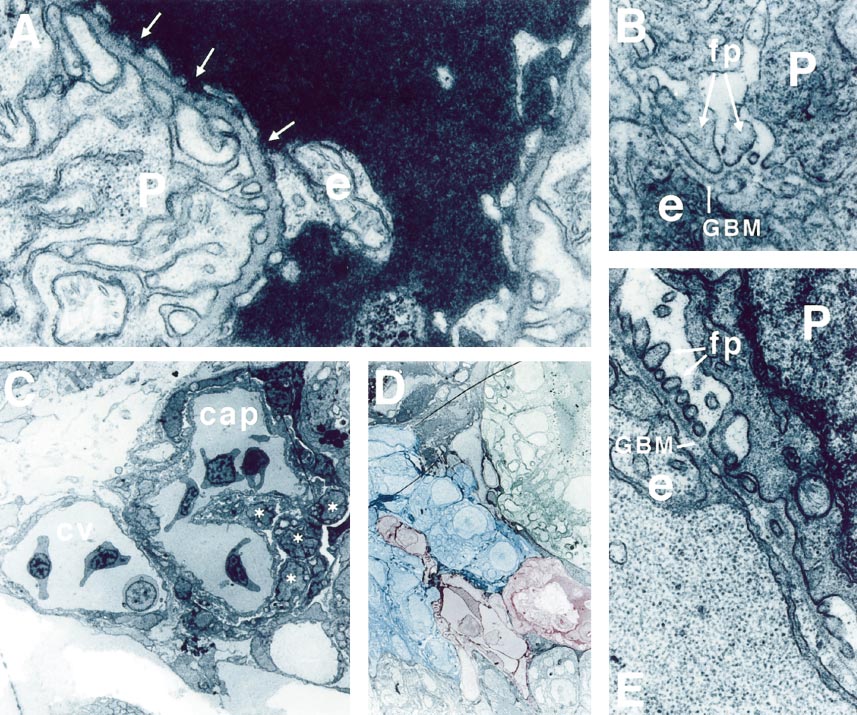
flh glomeruli contain appropriate glomerular cell types. In wildtype (A, B), podocytes (P) and endothelial cells (e) are found on opposite surfaces of the trilaminar glomerular basement membrane (GBM). Podocytes make well-formed, evenly spaced foot processes. Capillary endothelia show a fenestrated membrane morphology and pores are present (arrows in A). In flh glomeruli (C, D, E), blood-cell-containing capillaries (cap) are found adjacent to the cardinal vein (cv). In D, a vascular continuity (shaded in red) between the capillary space and the cardinal vein is visible and is surrounded by podocytes (shaded in blue). Capillary endothelia are intimately associated with morphologically identifiable podocytes in flh (asterisks in C). Podocytes and endothelia are organized on opposite sides of a well-formed and continuous GBM with well-formed lamina densa and lamina rarae (E). Podocytes appear morphologically normal with well-formed foot processes (fp). Significantly, endothelial cell membranes are nonfenestrated and continuous. Pores are absent. Occasionally, the membranes are detached from the GBM. Original magnifications: (A, B, E) 40,000x (C, D) 1600x
Reprinted from Developmental Biology, 222(1), Majumdar, A. and Drummond, I.A., The zebrafish floating head mutant demonstrates podocytes play an important role in directing glomerular differentiation, 147-157, Copyright (2000) with permission from Elsevier. Full text @ Dev. Biol.