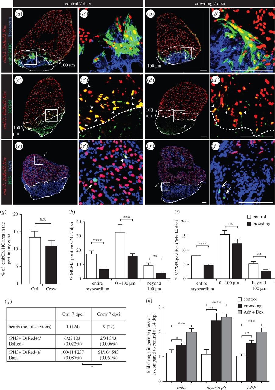
Fig. 3
Daily acute stress exposure after cryoinjury affects CM proliferation and triggers compensatory mechanisms associated with cardiac overload. (a-f) Immunofluorescence staining of heart sections at 7 dpci of cmlc2::DsRed2-Nuc (cardiac nuclei, red) transgenic zebrafish of control and stressed animals. Dashed lines encircle the cryoinjured parts and a 100 Ám-wide margin of the remaining myocardium along the injury border (peri-injury zone). (a-b′) No change in expression of embCMHC (embryonic isoform of cardiac myosin heavy chain, MAb N2.261, green) and fibronectin (blue) is observed between both experimental groups, N ≥ 4. (c-d′) The G1/S phase marker MCM5 (green) is reduced in CM nuclei of the stressed animals, particularly in the peri-injury zone. Arrowheads indicate double positive cells. (e-f′) The mitosis marker PH3 (green) is less frequent in the stressed animals as compared to controls. Arrows indicate mitotic non-CMs. (g-j) Quantification of immunofluorescence analysis shown in the representative images. N ≥ 8 hearts; three sections per heart. (k) qRT-PCR experiments revealed that exposure to crowding or dexamethasone/adrenaline (Adr + Dex) for 1 h d-1 during 14 days after cryoinjury results in upregulation of genes associated with cardiac compensatory response; N ≥ 2 sets, 10 hearts each. *p < 0.05, **p < 0.01, ***p < 0.001, ****p < 0.0001. Scale bars, 100 Ám.

