Image
Figure Caption
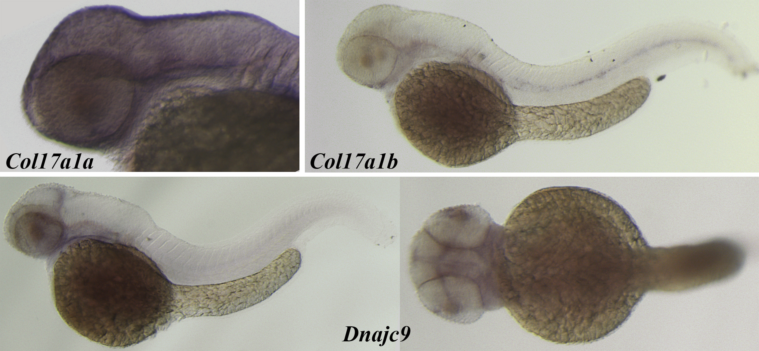
Fig. 6
Photomicrographs obtained after whole-mount in situ hybridization was performed using antisense probes to the Col17a1a, Col17a1b, and Dnajc9 genes on embryos 50 hours after fertilization and visualized with alkaline phosphatase?mediated staining. Col17a1a is observed in a punctate pattern on the epithelium, including over the developing cornea. Col17a1b transcripts are evident in the neuromast cells. Diffuse staining for Dnajc9 transcript was observed in the head region and retinal proliferative zone of the zebrafish embryos (lateral and dorsal views shown).
Figure Data
Acknowledgments
This image is the copyrighted work of the attributed author or publisher, and
ZFIN has permission only to display this image to its users.
Additional permissions should be obtained from the applicable author or publisher of the image.
Full text @ Ophthalmology

