
|
Fig. 4
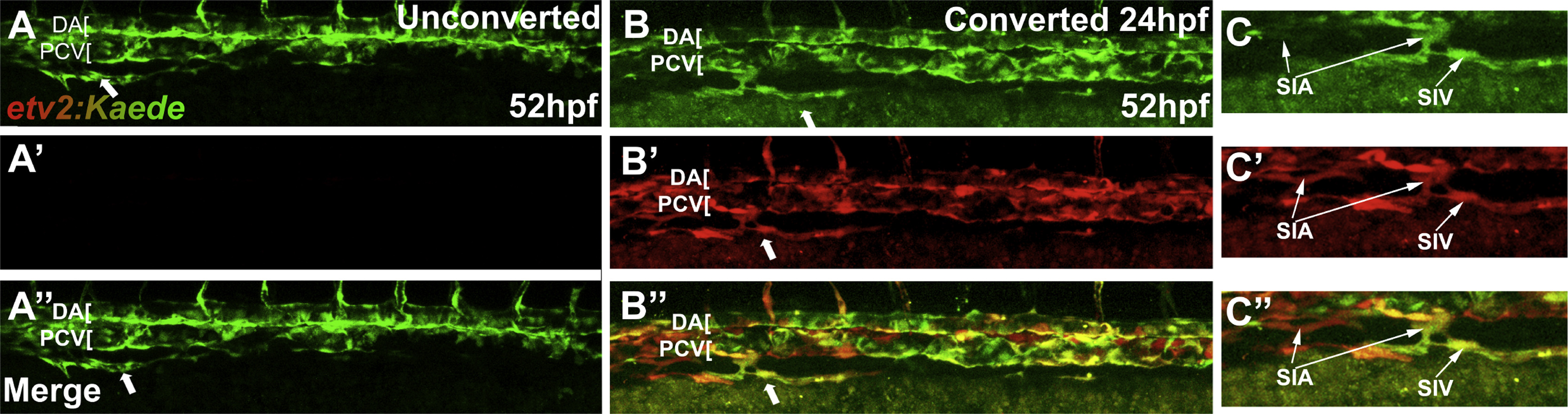
Cells in the sub-intestinal vein are derived from existing vasculature. Confocal microscope images of live Tg(etv2:Kaede) embryos following photoconversion. Note that etv2:Kaede line exhibits mosaic expression and not all vascular endothelial cells are labeled. (A-A′′) Unconverted control demonstrates that only converted cells are visible in the red channel. (B-B′′) Photoconversion at 24 hpf prior to any formation of intestinal vasculature results in all Kaede-positive cells in the SIV being marked by the converted (red) form of Kaede. This argues that SIV progenitors are derived from existing endothelial cells already expressing etv2:Kaede in the embryo at 24 hpf. (C-C′′) Cropped regions of converted embryos demonstrate that both the SIA and SIV are visible in the red channel. Arrows indicate SIV. Anterior-left, dorsal-top.
Reprinted from Developmental Biology, 411(1), Koenig, A.L., Baltrunaite, K., Bower, N.I., Rossi, A., Stainier, D.Y., Hogan, B.M., Sumanas, S., Vegfa signaling promotes zebrafish intestinal vasculature development through endothelial cell migration from the posterior cardinal vein, 115-27, Copyright (2016) with permission from Elsevier. Full text @ Dev. Biol.