IMAGE

Fig. 2

ID
ZDB-IMAGE-160324-2
Source
Figures for Zhang et al., 2016
Image
Figure Caption

Fig. 2

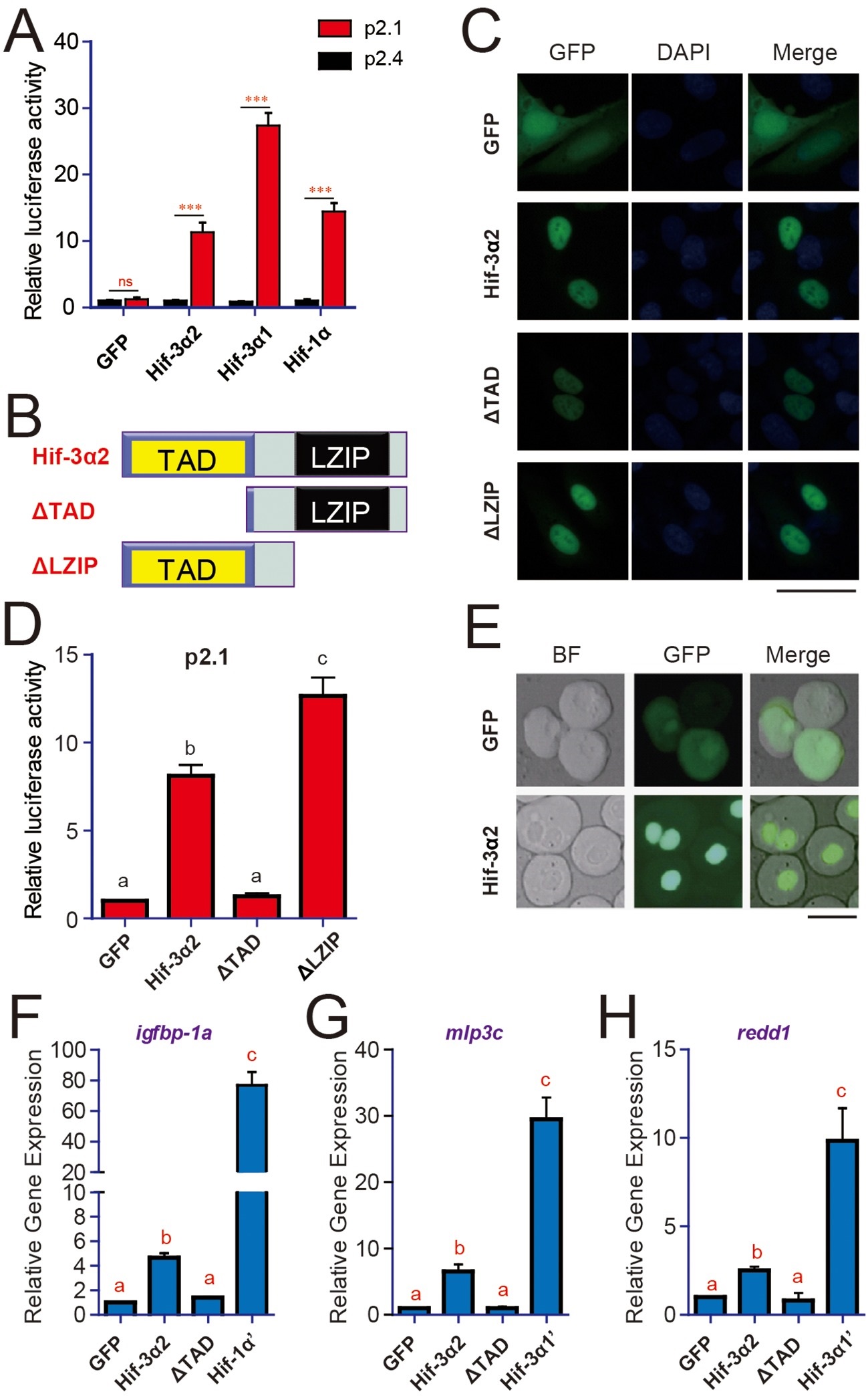
Hif-3α2 is a nuclear protein and has HRE-dependent transcriptional activity.(A) HRE-dependent transcriptional activity. HEK293 cells were transfected with the indicated plasmid together with 100 ng p2.1 (red) or p2.4 plasmid (black). The results are normalized and expressed as fold change over the GFP p2.4 group. Values are means S.E. (n = 3). ***p < 0.001. (B) Schematic diagram of the Hif-3α2 truncation mutants tested. (C) U2OS cells were transfected with the constructs shown in (B). The GFP signal was visualized 24 hr after transfection (left panels). Cells were counterstained with DAPI (middle panels). Merged views are shown in the right panels. Scale bar = 50 µm. (D) HRE-dependent transcriptional activity of Hif-3α2 mutants. Values are means + S.E. (n = 3). Groups labeled with different letters are significantly different from each other (P < 0.05). (E) Nuclear localization of Hif-3α2-EGFP in zebrafish embryo cells. Embryos injected with EGFP (600 pg) or Hif-3α2-EGFP capped mRNA (600 pg) were raised to 6 hpf under normoxia. The cells were dispersed and observed under bright field (BF) and GFP fluorescence microscopy. Scale bar = 50 µm. (F-H) Effects of Hif-3α2 (600 pg), Hif-3α1′ (stabilized Hif-3α1, 800 pg), and Hif-1α′ (stabilized Hif-1α, 800 pg) on endogenous gene expression. Embryos injected with the indicated capped mRNA were raised to 12 hpf under normoxia. The mRNA levels of the indicated genes were determined by qRT-PCR and normalized by the β-actin levels. Values are means S.E. (n = 3). Groups labeled with different letters are significantly different from each other (P < 0.05).

Acknowledgments
This image is the copyrighted work of the attributed author or publisher, and ZFIN has permission only to display this image to its users. Additional permissions should be obtained from the applicable author or publisher of the image. Full text @ Elife