
|
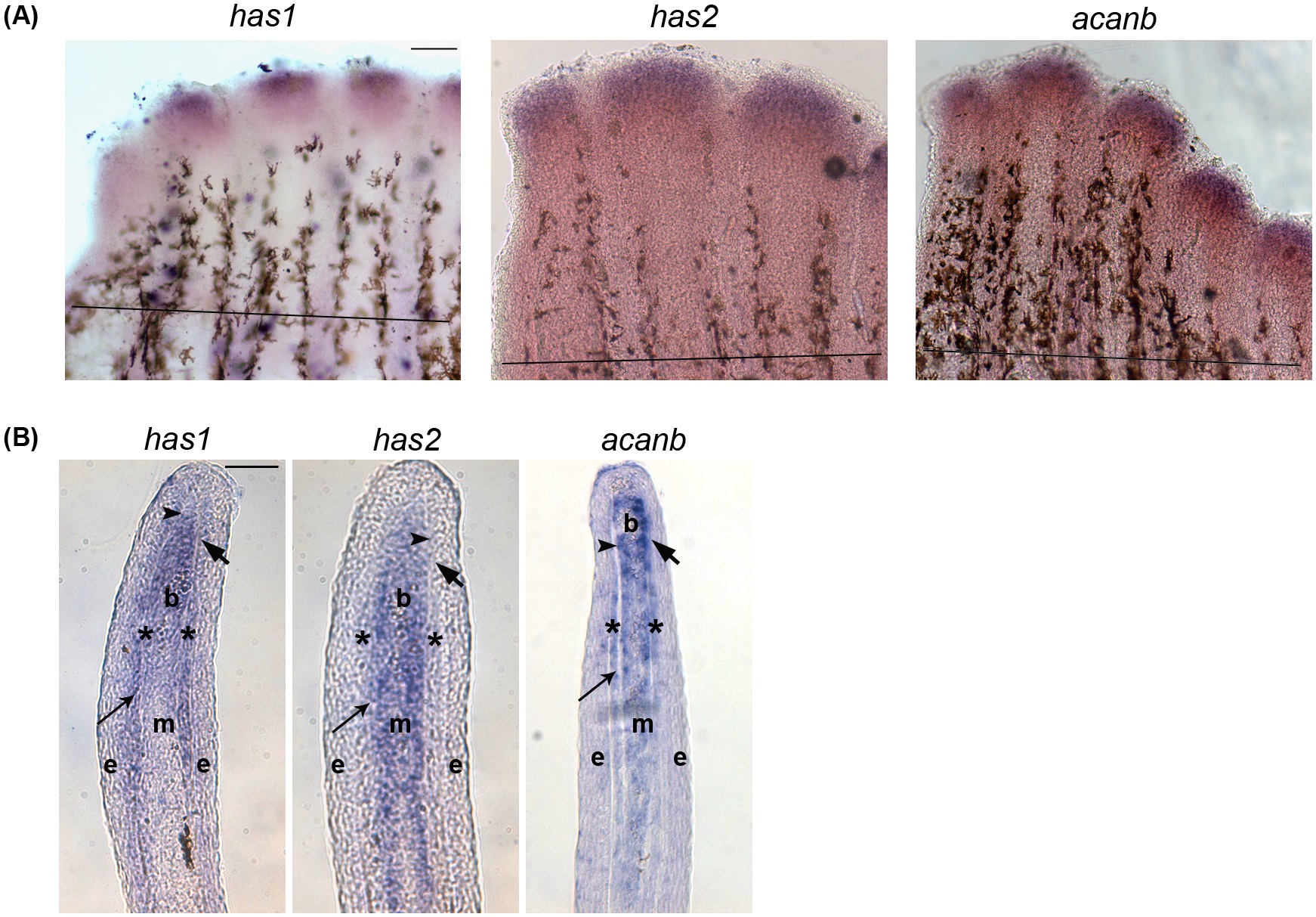
Fig. 1
In situ hybridization showing the expression of Hapln1a-ECM components on 5 dpa regenerating fins.
(A) Whole mount in situ hybridization of components of the Hapln1a-ECM. The genes has1, has2, and acanb are expressed during fin regeneration. The amputation plane is indicated by a black line in all the panels. Scale bar represents 100�m. (B) In situ hybridization on a WT 5 dpa cryo-section reveals compartmental expression of has1, has2, and acanb. Blastema (b), mesenchyme (m), and skeletal precursor cells (*). The thick arrow identifies the basal layer of the epidermis, which underlies the epidermis (e). The thin arrow identifies lepidotrichia and the arrowhead identifies the actinotrichia. Scale bar represents 50�m.