
|
Fig. 5
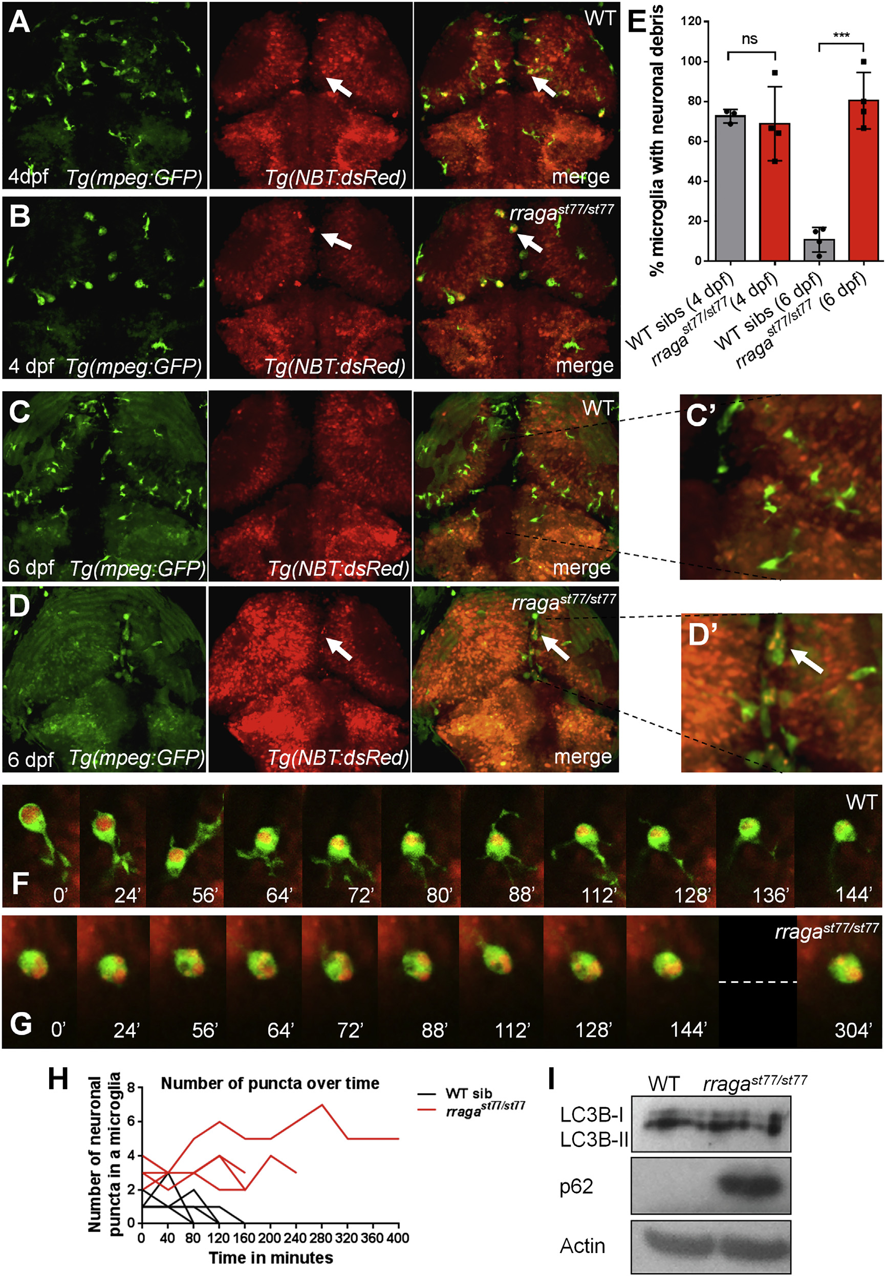
Accumulation of Undigested Neuronal Material in Microglia of In rragast77/st77 Mutants
(A?D, G, and H) Confocal images show living larvae bearing transgenes that label neurons Tg(nbt:DsRed) and microglia Tg(mpeg1:EGFP).
(A?E) In WT (A and C) and rragast77/st77 mutant (B and D) larvae, arrows indicate microglia that contain neuronal material. In WT larvae, more microglia contain neuronal material at 4 dpf (A and E) than at 6 dpf (C, C′, and E), whereas most microglia in mutants contained neuronal material at both 4 and 6 dpf (B, D, D′, and E). Each point in (E) represents the data from one larva.
(F and G) Frames from time-lapse movies of WT (F) and rragast77/st77 (G) mutant larvae starting at 4 dpf, with time points indicated in minutes.
(H) Quantification of the number of neuronal puncta inside microglia in time-lapse movies of WT and rragast77/st77 mutant larvae. Each line represents a single microglia (WT, n = 4 cells from three larvae; mutant, n = 4 cells from three larvae).
(I) Immunoblot of whole-animal protein lysates at 5 dpf shows normal LC3-I to LC3-II conversion, but an accumulation of p62 in rragast77/st77 mutants. Actin is shown as a loading control.
All larvae shown in (A)?(H) were genotyped by PCR after photography. Larvae analyzed in the immunoblot (I) were genotyped as described in the Experimental Procedures.