
|
Fig. 3
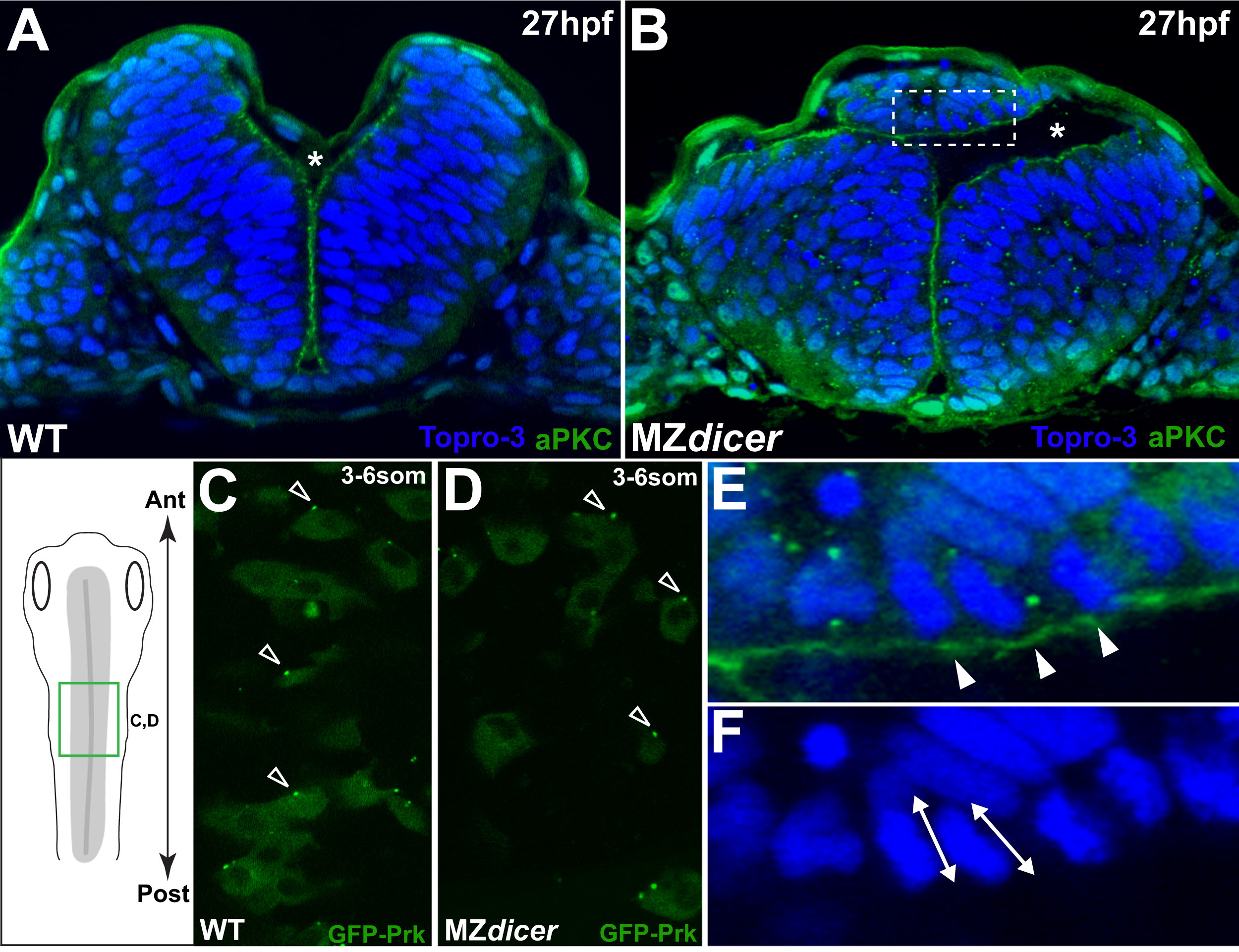
Ectopic MZdicer neural progenitors retain apicobasal and PCP polarity. (A and B) Transverse sections at 27hpf. Topro3, nuclear stain. At this stage, lumen inflation has initiated. Asterisk indicates luminal space. (B) MZdicer embryo with a dorsal ectopic cell aggregate. (E) Inset of panel B; arrowheads indicate aPKC (green) localization to apical membrane. (F) Inset of panel B; double arrows indicate elongated nuclear shape characteristic of polarized epithelial cells. (C and D) Dorsal view (green box in schematic) of GFP-Prickle scatter labeling. GFP-Prk localizes to the anterior edge (arrowheads) of (C) wildtype neural cells (71/94; 76%) and (D) MZdicer mutant cells (39/49; 80%). Anterior to top. (For interpretation of the references to color in this figure legend, the reader is referred to the web version of this article.)
Reprinted from Developmental Biology, 409(2), Takacs, C.M., Giraldez, A.J., miR-430 regulates oriented cell division during neural tube development in zebrafish, 442-50, Copyright (2016) with permission from Elsevier. Full text @ Dev. Biol.