Fig. 3
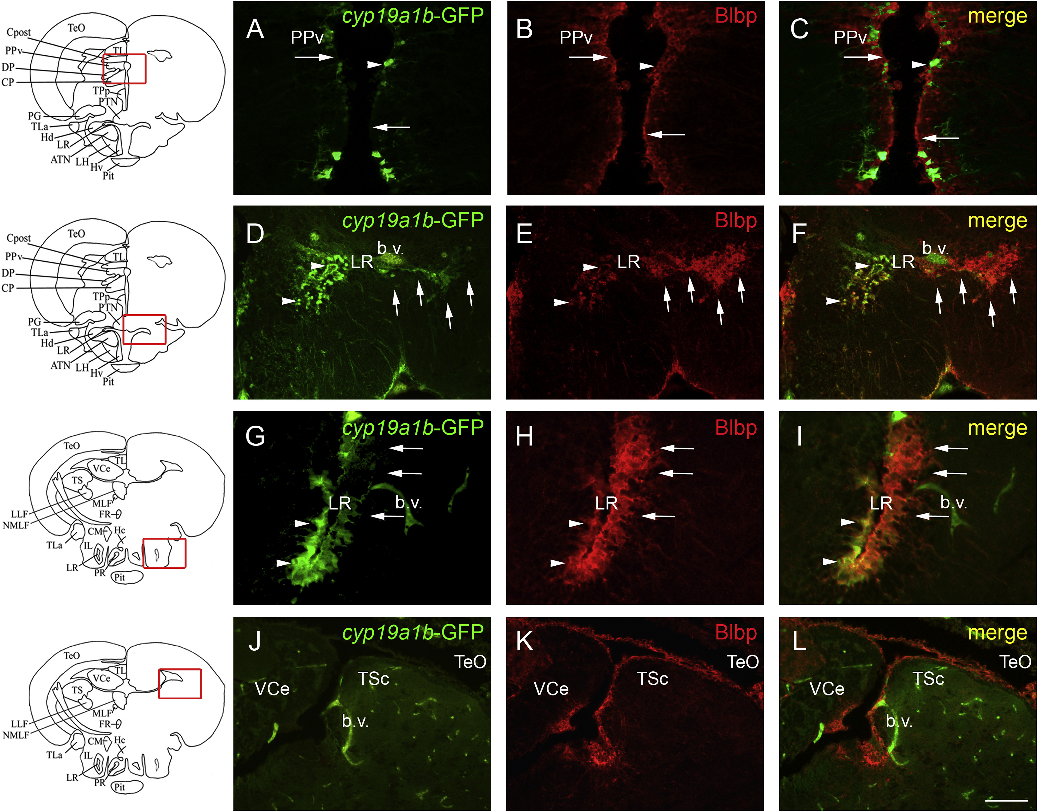
cyp19a1b-GFP and Blbp show differences of expression in more caudal regions of the brain of adult zebrafish. A-L: Blbp (red) immunohistochemistry on cyp19a1b-GFP (green) transgenic zebrafish line show discrepancies in expression of AroB and Blbp markers. In the pretectal periventricular nucleus, radial glia mainly expresses Blbp while the transgene is only expressed in few cells (A-C). In the dorsal zone of the periventricular hypothalamus, radial glial cells surrounding the lateral recess of the hypothalamus exhibit heterogeneous expression of both markers (D-I). When the lateral recess starts to open (D-F), numerous radial glial cell express Blbp along the LR, while GFP is rarely expressed. In more caudal sections, radial glial cells of the medial part of the LR nucleus exhibit GFP and Blbp staining (arrowheads in G-I), and the ?external? ones mainly express Blbp and not GFP (arrows in G-I). Blbp staining is observed around the torus semicircularis and in numerous radial glial cells from the optic tectum. In contrast, GFP is almost not detected (J-L). Bar: 25 Ám (G-I); 50 Ám (A-F and J-L).
Reprinted from Gene expression patterns : GEP, 20(1), Diotel, N., Vaillant, C., Kah, O., Pellegrini, E., Mapping of Brain lipid binding protein (Blbp) in the brain of adult zebrafish, co-expression with aromatase B and links with proliferation, 42-54, Copyright (2016) with permission from Elsevier. Full text @ Gene Expr. Patterns

