
|
Fig. 3
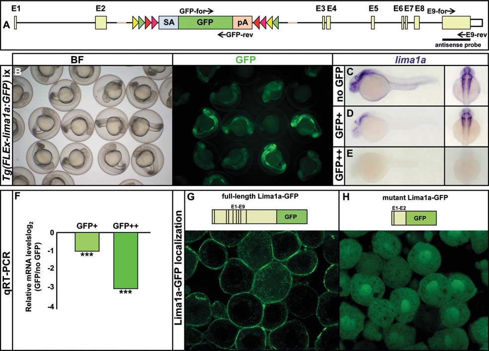
Functional consequences of the integration of SAGFLEx into the lima1a locus. (A) Schematic representation of SAGFLEx integrated into intron 2 of the lima1a locus in Tg(FLEx-lima1a:GFP). The positions of the primers used for qRT-PCR analysis (GFP-for/GFP-rev, E9-for/E9-rev) and the lima1a exon 9 probe are indicated. (B) Mating of heterozygous Tg(FLEx-lima1a:GFP) animals results in offspring with no, medium and strong GFP fluorescence. (C–E) Expression of lima1a in embryos with no, medium and strong GFP fluorescence (no GFP, GFP+, GFP++) using an exon 9 probe. Lateral and dorsal views at 28 hpf. (F) qRT-PCR analysis of lima1a exon 9 expression in embryos with medium and strong GFP fluorescence compared to siblings with no GFP fluorescence. ***P value ≥ 0,001. (G, H) Cellular localization of GFP fusions with wildtype Lima1a encoded by exons 1–9 (E1–E9) and truncated Lima1a encoded by exons 1–2 (E1–E2).