
|
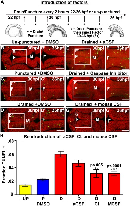
Fig. 3
Introduction of Caspase 3 inhibitor and mouse CSF but not aCSF prevents cell death. (A) Experimental design. Drainage/puncture occurred every 2 hours from 22 to 36 hpf. From 30 to 36 hpf a factor was injected every 2 hours into the brain ventricles after drainage/puncture. Embryos were assayed at 36 hpf. (B–G) Dorsal view of TUNEL (green) and propidium iodide (red) in un-punctured (B) and punctured (C) embryos injected with DMSO or drained embryos injected with DMSO (D) aCSF (E), Caspase 3 inhibitor (F), or E10.5 mouse CSF (G). (H) Quantification of TUNEL. Data represented as mean ± SEM. F = forebrain, M = midbrain, UP = unpunctured, P = punctured, D = drained, CI = Caspase 3 inhibitor, MCSF = E10.5 mouse CSF. Scale bars = 50 µm.