
|
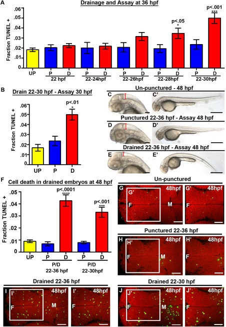
Fig. 2
CSF is required from 25 to 30 hpf, while cell death persists at 48 hpf. (A) Quantification of TUNEL. X axis indicates puncture/drainage period. All assayed at 36 hpf. (B) Quantification of TUNEL at 30 hpf after puncture/drainage from 22 to 30 hpf. (C–E) Brightfield dorsal and lateral (C′–E′) images of 48 hpf embryos after puncture/drainage from 22 to 36 hpf. (F) Quantification of TUNEL at 48 hpf. (G–J) Dorsal view of TUNEL (green) and propidium iodide (red) in 48 hpf embryos after puncture/drainage from either 22–36 hpf (H–I) or 22–30 hpf (J) or unpunctured (G). Data represented as mean ± SEM. F = forebrain, M = midbrain. UP = unpunctured, P = punctured, D = drained. Scale bars = 50 µm.