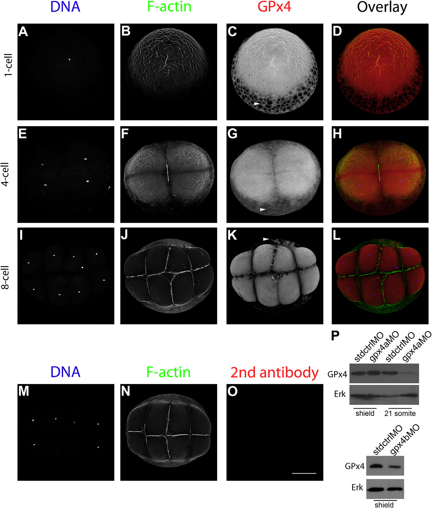
Fig. 2
GPx4 immunofluorescence localization patterns in whole zebrafish embryos from 1- to 8-cell stages as detected by laser confocal microscopy. 1-cell embryo (A, B, C and D). 4-cell stage embryo (E, F, G and H). 8-cell stage embryos (I, J, K and L). 8-cell stage control embryo in which the primary antibody was omitted but was incubated in the presence of secondary antibody (M, N and O). DNA, Hoechst-stained embryos (A, E. I and M). F-actin, phalloidin Alexa 488 stained embryos (B, F, J and N). GPx4, immunolocalization in whole embryos (C, G and K). Arrowheads indicate GPx4-positive signals in the yolk (C, G and K). Animal pole views are shown. Scale bar 200 Ám. P, GPx4 antibody validation in zebrafish embryo samples at the shield stage. Gpx4a and b were down regulated by morpholino injection. A signal decrease was verified by western blotting, in which we compared immunoblot results with total protein samples from gpx4a or gpx4b morpholino injected embryos against standard control morpholino injected embryos. After immunoblotting for GPx4, the same membranes were stripped and immunoblotted against total ERK to confirm that the same amount of protein was loaded into the wells.
Reprinted from Gene expression patterns : GEP, 19(1-2), Mendieta-Serrano, M.A., Schnabel-Peraza, D., LomelÝ, H., Salas-Vidal, E., Spatial and temporal expression of zebrafish glutathione peroxidase 4 a and b genes during early embryo development, 98-107, Copyright (2015) with permission from Elsevier. Full text @ Gene Expr. Patterns

