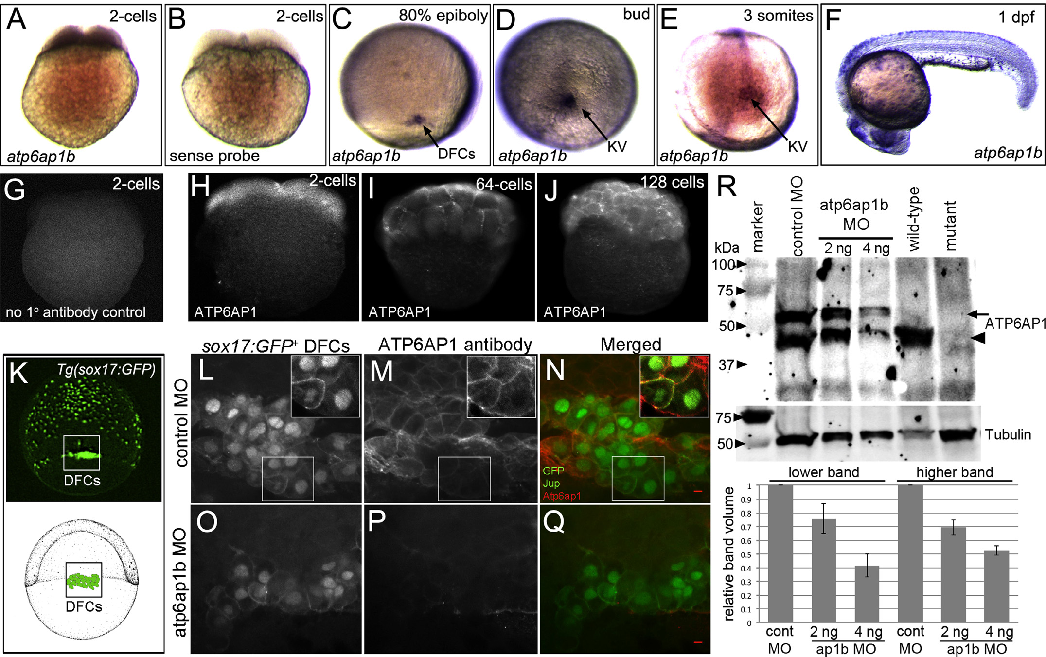
Fig. 1
Atp6ap1b is maternally supplied and prominently expressed in dorsal forerunner cells and Kupffer?s vesicle. (A?F) RNA in situ hybridizations of atp6ap1b during early zebrafish development. (A) Antisense atp6ap1b probes revealed maternal atp6ap1b mRNA in the 2-cell embryo. (B) A control atp6ap1b sense probe showed no staining. (C?E) atp6ap1b mRNA was detected in DFCs and KV cells during epiboly and early somite stages. (F) atp6ap1b was observed in mucus secreting cells, eye and brain at 24 hpf. (G?J) Fluorescent immunostaining with ATP6AP1 antibodies detected maternal protein at the 2-cell (H), 64-cell (I) and 128-cell (J) stages. There was no signal in control experiments that lacked ATP6AP1 antibody (G). (K) Schematic diagram and whole-embryo view of a Tg(sox17:GFP) embryo that expresses GFP in DFCs and endoderm at 80% epiboly. The box indicates the approximate region of interest shown in L?Q. (L?Q) ATP6AP1 antibodies labeled all cells during epiboly stages, including GFP+ DFCs, in control MO injected Tg(sox17:GFP) embryos (L?N). Jup antibodies were used to mark plasma membranes in DFCs. Insets show ATP6AP1 staining was observed in the cytoplasm and plasma membranes. ATP6AP1 staining was reduced in Atp6ap1b MO embryos (O?Q). (R) Western blot analysis of Atp6ap1 expression. ATP6AP1 antibodies detected prominent bands just under 50 kDa (arrowhead) and just higher that 50 kDa (arrow) in control embryos at the bud stage (10 hpf) that were reduced in a dose-dependent fashion in atp6ap1b MO embryos. The lower band was detected in wild-type embryos at 7 dpf, and was reduced in atp6ap1ba82/a82 mutants. Tubulin was used as a loading control. The graph shows average relative intensities of both Atp6ap1 bands from three MO experiments.
Reprinted from Developmental Biology, 407(1), Gokey, J.J., Dasgupta, A., Amack, J.D., The V-ATPase accessory protein Atp6ap1b mediates dorsal forerunner cell proliferation and left-right asymmetry in zebrafish, 115-30, Copyright (2015) with permission from Elsevier. Full text @ Dev. Biol.

