Image
Figure Caption
Fig. S3
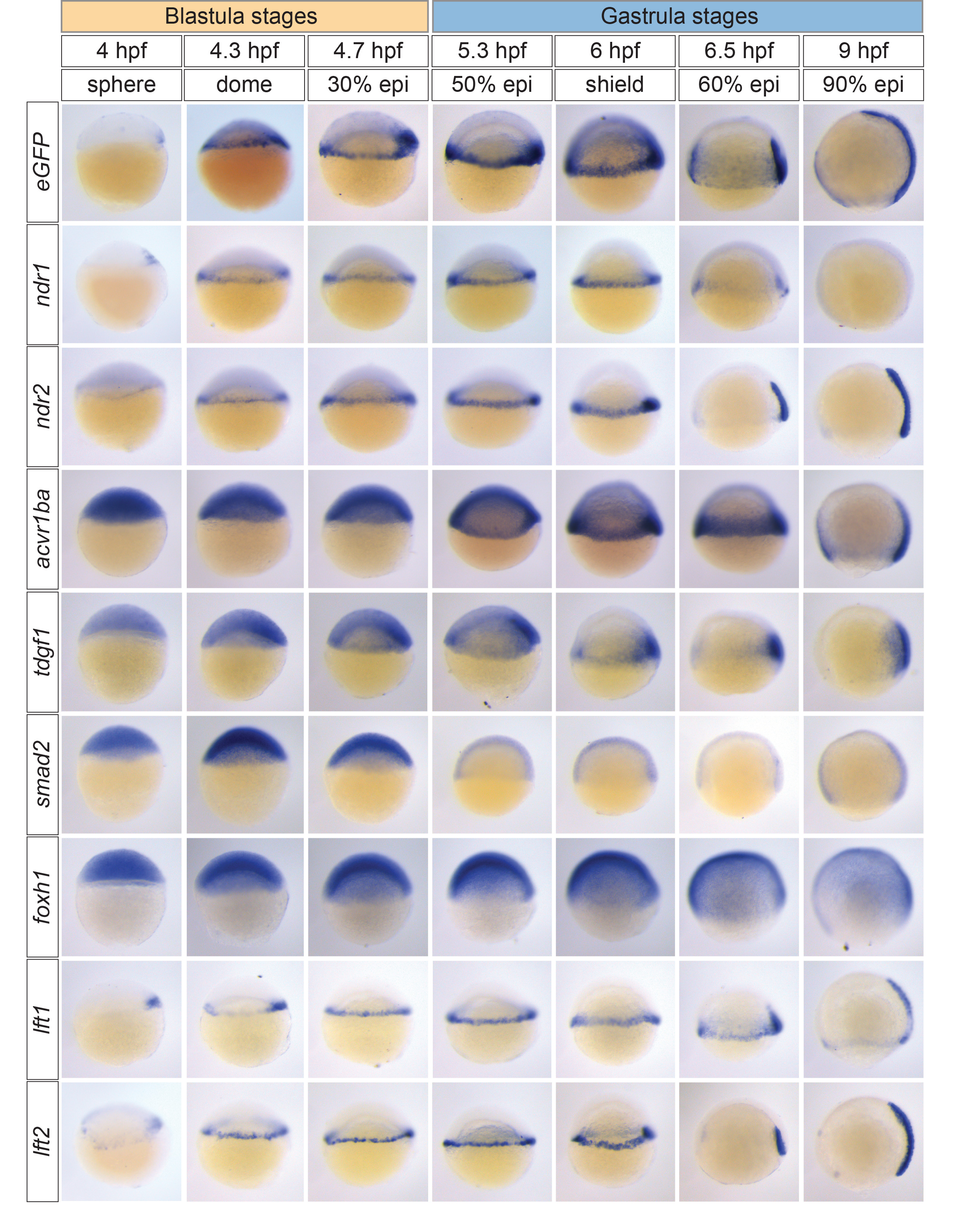
Comparison of Nodal signaling activation in Tg(ARE:eGFP) embryos and expression of core components of the Nodal signaling pathway
WISH for eGFP, ndr1 (sqt), ndr2 (cyc), acvr1ba (tar-a), tdgf1 (oep), smad2, foxh1 (sur), lft1 and lft2 in blastula and gastrula stage Tg(ARE:eGFP) embryos.
Acknowledgments
This image is the copyrighted work of the attributed author or publisher, and
ZFIN has permission only to display this image to its users.
Additional permissions should be obtained from the applicable author or publisher of the image.
Reprinted from Developmental Cell, 35, van Boxtel, A.L., Chesebro, J.E., Heliot, C., Ramel, M.C., Stone, R.K., Hill, C.S., A Temporal Window for Signal Activation Dictates the Dimensions of a Nodal Signaling Domain, 175-185, Copyright (2015) with permission from Elsevier. Full text @ Dev. Cell

